NEET Biology Question Bank - Neural Control and Coordinations in Animals
Finished viewing? Save it for later —
Download NEET Biology Question Bank - Neural Control and Coordinations in Animals (PDF · 25 pages)About NEET Biology Question Bank - Neural Control and Coordinations in Animals
NEET Biology Question Bank - Neural Control and Coordinations in Animals is available here for free download. Published by NTA for National Eligibility cum Entrance Test (Undergraduate), this question bank can be viewed online or downloaded as a PDF (25 pages). Candidates preparing for National Eligibility cum Entrance Test (Undergraduate) can use NEET Biology Question Bank - Neural Control and Coordinations in Animals to understand the exam pattern, the type of questions asked, and the overall difficulty level.
Frequently Asked Questions
How can I download NEET Biology Question Bank - Neural Control and Coordinations in Animals?
Open this page and click the Download button to save NEET Biology Question Bank - Neural Control and Coordinations in Animals as a PDF. It is completely free on AglaSem Docs.
Is NEET Biology Question Bank - Neural Control and Coordinations in Animals free to download?
Yes. NEET Biology Question Bank - Neural Control and Coordinations in Animals can be viewed online and downloaded as a PDF free of cost on AglaSem Docs.
How many pages does NEET Biology Question Bank - Neural Control and Coordinations in Animals have?
NEET Biology Question Bank - Neural Control and Coordinations in Animals contains 25 pages, which you can read online or download together as a single PDF.
Where can I find more National Eligibility cum Entrance Test (Undergraduate) study material?
You can find more National Eligibility cum Entrance Test (Undergraduate) question papers, sample papers, syllabus, and answer keys on AglaSem Docs.
NEET Biology Question Bank - Neural Control and Coordinations in Animals – Text
Read the full text of this question bank below — useful to quickly search, copy and reference the content online without downloading the PDF.
📄 View text version (25 pages)
Page 1
l Nervous System :
- In Human being all physiological processes control and coordination by Nervous system and Endocrine
system.
- The nervous system provides and organised network of nerves for fast coordination.
- The endocrine system provides chemical integration through hormones.
- Nerve cell is the structural and functional unit of the nervous system.
- Three main functions of nervous system : (1) Receiving sensory stimuli from external and internal
environment by nerves to the brain. (2) Processing the stimuli informations by brain. (3) Responding
to stimuli transrmitting impulses from brain to body parts of cells.
- The nervous system of hydra is composed of network of nerve cells.
- The nervous system is well developed in insects, which consists of brain, ganglia and nervous system.
- Vertebrates have a highly developed nervous system.
l Structures of Nervecell : (Three parts)
(1) Cyton : The cyton may be oval, rounded or star shaped neuroplasma, which has a relatively large
and spherical nucleus. Neuroplasm has mitochondria, golgibody and small basophilic granules i.e. Nissl's
grenules.
(2) Dendrites : Several short branched tapering afferent processes.
(3) Axon : A Single, cylindrical and very long efferent processes with synaptic knob.
l Synapse : An ending branch of axon of first neuron and sequincial dendrites of record neuron
never directly attached with each other but very small physical space is there so it is called synapse.
l On basis of processes types of neurons :
(1) Unipolar neuron : One axon only e.g. found in embryonic stage.
(2) Bipolar neuron : One axon and one dendrite. eg. found in retina of eye.
(3) Multipolar neuron : One axon and more dendrites present in the cerebral cortex.
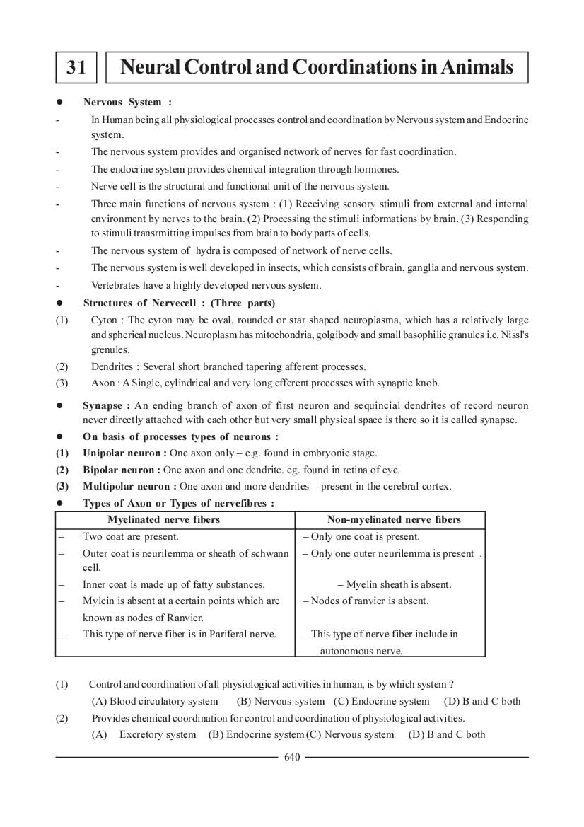
l Types of Axon or Types of nervefibres :
Myelinated nerve fibers Non-myelinated nerve fibers
Two coat are present. Only one coat is present.
Outer coat is neurilemma or sheath of schwann Only one outer neurilemma is present .
cell.
Inner coat is made up of fatty substances. Myelin sheath is absent.
Mylein is absent at a certain points which are Nodes of ranvier is absent.
known as nodes of Ranvier.
This type of nerve fiber is in Pariferal nerve. This type of nerve fiber include in
autonomous nerve.
(1) Control and coordination of all physiological activities in human, is by which system ?
(A) Blood circulatory system (B) Nervous system (C) Endocrine system (D) B and C both
(2) Provides chemical coordination for control and coordination of physiological activities.
(A) Excretory system (B) Endocrine system (C) Nervous system (D) B and C both
640
Page 2
(A) Nervous system (B) Endocrine system (C) Blood vascular system (D) A and B both
(4) In which animal phylum nerve net type nervous system is present ?
(A) Porifera (B) Coelenterata (C) Protozoa (D) Arthropoda
(5) Cyton is which shape ?
(A) Star (B) Oval (C) Round (D) All
(6) Where is secretory vesicle, located in nerve cell ?
(A) Cyton (B) Synapse (C) Synaptic knob (D) Nerve fibre
(7) Location of neuron, having one axon ......... .
(A) cerebral cortex (B) retina (C) embryonic stage (D) all
(8) Location of neuron having one dendron.
(A) Embryonic (B) Cerebral cortex (C) Retina (D) All of above
(9) Type of neuron having more than two axons.
(A) Multipolar (B) Bipolar (C) Unipolar (D) None
(10) Nerve, which has covering of schwann's cell.
(A) cranial nerve (B) Spinal nerve
(C) Autonomous nerve (D) All of above
(11) Nerve with two membranes surrounding nerve fibre ......... .
(A) cranial nerve (B) autonomous nerve
(C) spinal nerve (D) A and C
Answers Ñ (1-D), (2-B), (3-A), (4-B), (5-D), (6-C), (7-C), (8-C), (9-A), (10-D), (11-D)
l Structures of nerve fiber resting potential and Formation of nerve impulse :
l Structure of nerve fiber : The neuroplasm possesses a higher negative charge compared to it,
there is a higher positive charge on the outerside of plasma membrane. The distribution of
positive and negative ions is responsible for this differences in electrical charges.
- This difference in electrical charges between the inside and the outisde plasma membrane is
called the 'membrane potential'. In an unexcited state of nerve fibre, it is called 'resting potential' !
It can be measured using electrodes and voltmeter. Plasma membrane of nerve fiber has main
two parts.
(1) Bilayer lipids - which is impermeable for ions.
(2) Specific protein present in coat.
Functions :
(1) Ion channels : Aqueous and made up of protein - carry out two way transport of ions
selectively. - permeable to any one kind of ion only - Na+, K+, Cl- and Ca+2 ion channels can occur
transport in direction of concentration gradiant- Ion channels can be opened and closed.
Chemical and electrical charges are responsible for this - bring differences in electrical potential.
(2) It play key roll to maintain the electrical potential and change.
(3) Ion pumps : Transportation of ions opposite to concentration gradient - In transportation energy
consume as ATP - Mainly Na+ and K+ pump is there. - By the Na+ pump 2 Na+ goes outside.
While 3K+ comes inside by K+ pump - Ion pump maintain potential.
641
Page 3
side and a positive electrical charge occurs on its outerside.
l Such a nerve fibre is called polarized.
- For this, ions responsible Na+ are concentrated on the outside of plasma membrane whereas K+
are concentracted on its inside.
- Moreover protein molecules having negative charges on them also occur. They can not move out
of plasma membrane.
l Active potential or initiation of the Impulse :
- Nerve fibre can become excited in verious ways. Touch, smell, pressure, chemical changes etc.
include this.
- A change in the polarity of nerve fibre is called active potential.
- In the excited region Na + channels open up; the electrical excitation generated in plasma
membrane in this region is reponsible for this. Na + ions are poured inside through its ion
channels in plasma membrane. The lower concentration of Na + inside is responsible for
this. Moreover, negatively charged proteins are also responsible. Thus, due to a sudden influx of
a large number of Na + towards the inside, the plasma membrane in this region becomes
positively charged on its inside. This phenomenon is called- depolarization. It is called active potential.
- It lasts for a very short period i.e a millionth part of a second (nearly 0.5 milli sec). The excited
region immediately becomes repolarised.
- For repolarization, the process of closure of Na+ ion channels is responsible, simultaneously, K+
ion channels open up and K+ ions go out of the plasma membrane.
- At the end of this phase, a difference in concentration of positive ions on two sides of plasma
membrane is generated. Concentration of Na+ increases on inside and that of K+ increases on the outside.
- The activity of Na+ - K+ pump removes this imbalance.
(12) Conduction of nerve impulse by nerve fibre is which type of process ?
(A) electrical (B) chemical (C) electrochemical (D) physical
(13) Which electrical charge is more towards outerside of neurolemma of inactive nervefibre ?
(A) Negative (B) Positive (C) Negative and positive (D) Neutral
(14) Membrane potential is ......... .
(A) difference of electric charge, innerside of nerve cell
(B) difference of electric charge, on outer surface of nerve cell
(C) difference of outer and inner electric charge of nerve cell
(D) inner and outer electric charge of nerve cell, become similar
(15) Conduction of ions by Na+ - K+ pump is of ........ type.
(A) Antiport (B) Symport (C) Uniport (D) biport
(16) Protein which conducts ions without utilization of ATP in neurilemma of nerve cell.
(A) Na+ - K+ pump (B) Na+ passage (C) K+ passage (D) B and C
(17) ............. is responsible for resting membrane potential of nerve fibre.
(A) Na+ channel (B) K+ channel (C) Na+ - K+ channel (D) Na+- K+ pump
(18) .......... is responsible for changing resting membrane potential of nerve fibre.
(A) Na+ channel (B) K+ channel (C) Na+ and K+ channel (D) Na+- K+ pump
(19) What is duration of active membrane potential on nerve fibre ?
(A) 1 Sec (B) 0.5 Millisec (C) 2 Sec (D) 5 Sec
(20) Which ions are responsible for creating active membrane potential ?
(A) K+ (B) Ca++ (C) Cl- (D) Na+
642
Page 4
(A) Na+ - K+ channels closed (B) Na+ - K+ channel open
(C) Na+ channel closed, K+ channel open (D) Na+ channel open, K+ channel closed
(22) Proper for repolarised nerve fibre is ......... .
(A) K+ more towards innerside (B) Na+ more towards innerside
(C) K+ more towards outerside (D) B and C
(23) Filled with water and formed of protein
(A) Na+channel (B) K+channel (C) Ca+2 channel (D) All of above
(24) Responsible for opening of Na+ channel of nervefibre.
(A) Touch (B) Smell (C) Pressure (D) All of above
(25) Important characteristic of active membrane potential ......... .
(A) Intensity of stimulation should constant (B) Intensity of stimulation should not constant
(C) Intensity of stimulation is specific and less (D) All of above
Answers : (12-C), (13-B), (14-C), (15-A), (16-D), (17-D), (18-C), (19-B), (20-D), (21-C), (22-D),
(23-D), (24-D), (25-C)
l Conduction of nerve impulse :
- The nerve impulse in nerve fibre conducted along one direction.
- This process is self-induced because when the membrane electric potential is reduced in the
nearby region, the ion-channels in the region automatically open up.
- Thus sequential depolarization and repolarization progress and the nerve impulse moves along in
that direction.
- To protect the impulse weakening through diffusion all arounds nerve fibre is surrounded by a
medullary layer. This conduction is very speedy (100 meters per second).
- In these nerve fibres, active potential after it is generated, does not move in cyclic waves
gradually. It moves directly from one Ranvier's node to the next node. Such a conductions is
known as 'Saltatory Conduction.'
l Transmission of Impulse by Synapse (Phases) :
Impuse - The synaptic knob of the axon, it depolarises the presynaptic membrane - Ca+2
permeability increases. - Ca+2 from the synaptic cleft enter into synaptic knob - Releases
acetylcholine in synapse- Acetylcholine receptors complex - Post synaptic membrane allows Na+
to enter the cell and cause depolarization - Generate new action potential in the post synaptic
membrane. Now the nerve impulse passes to next neuron. - Now acetylcholine is hydrolysed in
presence of acetylcholine esterase - Resynthesised into acetylcholine in presence of ATP.
- Ca+2 and Na+ ions presence in synapse - postsynaptic membrane having receptive protein.
- Acetyle choline esterase present in synaptic cleft and post synaptic membrane.
- In synapse direction of impulse is not proper because dendrites can not produce neurotransmitters.
643
Page 5
(A) depolarization and depolarization (B) repolarization and repolarization
(C) depolarization and repolarization (D) repolarization and depolarization
(27) Prevents spreading of nerve impulse intosurrounding medium ......... .
(A) nourilemma (B) myelin layer
(C) sarcolemma (D) both A and B
(28) Shows saltatory conduction of nerve impulse ......... .
(A) cranial nerve (B) spinal nerve
(C) autonomous nerve (D) A and B both
(29) In which nerve, active membrane potential once formed moves ahead cyclic, continuously without
interruption ......... .
(A) Sympathetic nerve (B) Parasympathetic nerve
(C) Spinal nerve (D) A and B
(30) Conduction of nerve impulse is possible through ......... .
(A) Acetic acid (B) Choline (C) Acetyl choline (D) Acetyl Co-A
(31) For conduction of nerve impulse through synapse ......... .
(A) Ca+2 should come out from presynaptic membrane
(B) Ca+2 should enter presynaptic membrane
(C) Ca+2 should enter post-synaptic membrane
(D) Ca+2 should come out through post-synaptic membrane
(32) When Na+ Channel of post synaptic membrane open up ?
(A) When Ca+ enters synaptic node.
(B) When acetyl choline released in synapse.
(C) When acetyl choline is hydrolysed.
(D) Acetyl choline receptor unit is formed.
(33) Where does hydrolysis of acetyl choline occur ?
(A) Synaptic node (B) Synaptic groove
(C) Post synaptic membrane (D) Pre-synaptic membrane
(34) True sequence for conduction of nerve impulse through synapse.
(1) Hydrolysis of acetylcholine. (2) Secretion of acetyl choline
(3) Entry of Ca++ into synaptic knob. (4) Formation of acetyl choline receptor complex.
(A) 2 - 3 - 4 - 5 - 1 (B) 5 - 3 - 4 - 2 - 1
(C) 3 - 2 - 4 - 5 - 1 (D) 1 - 2 - 3 - 4 - 5
(35) Location of Acetyl choline esterase - ......... .
(A) Synaptic knob (B) Pre synaptic membrane
(C) Post-synaptic membrane (D) All of above
Answers : (26-C), (27-B), (28-D), (29-D), (30-C), (31-B), (32-D), (33-B), (34-C), (35-C)
644
Page 6
Human Nervous System (parts)
¯
¯ ¯
Central Nervous System (CNS) Peripheral Nervous System(PNS)
¯ ¯
¯ ¯ ¯ ¯
Brain Spinal cord Cranial ® Sensory, ¬ Spinal
Nerves motor nerves
or mixed types
¯ ¯
Systemic Nervous System Auto nomous Nervous system (ANS)
¯ ¯ ¯
Conduction of nerve Sympathatic® Involuntary ¬ parasympathatic
impulsions toward Nervous System organs and Nervous
skeleton muscles (SNS) visceral muscles system(PNS)
toward coduct of
nerve impulse
l Central Nervous system : The CNS consists of the brain and the spinal cord. The CNS has
two distinct regions (1) White matter- White in colour and consists of myelienated fibres (2)
Gray matter- cyton bodies along with their dendrites and axon.
- Meaninges of Central Nervous System (CNS):
¯ ¯ ¯
(1) The inner membrane is (2) Middle membrane (3) The outer membrane is dura
pia matter which is thin arachnoid matter which matter. The thickest membrane
highly vascular membrane is a thin transparent mem- which covers the brain. It is
brane but non vascular also adherent to the inner
surface of cranial bones by
fibrous and vascular processes.
l Human brain :
- Weight about 1200 to 1400 gm.
- Number of neurons is about 100 billions.
- The brain can be divided into three major parts.
(1) Fore brain (2) Mid brain (3) Hind brain
(1) Fore brain :
A. Olfactory Lobes : Small, club shaped solid appear- only found in ventral view, covered by cerebral,
hemisphere.
B. Cerebrum : The largest parts of brain. - They are separated from each other by a longitudinal cerebral
fissure. Two hemispheres are connected by a large bundle of myelinated fibres known as corpus
callosum. Surface of the ecrebral cortex is highly folded to increase the area for accommodating more
nerve cells. The folds are called gyri and the depression between them are Known as Sulci Three wide
and deep sulci are termed as fissures. Which divide each hemisphere into four lobes.
(B1) Anterior frontal lobe: The premotor area fronted lobe controls involuntary movements and
autonomic nervous system. The associated area of the frontal lobe is concerned with memory,
reasoning,learning and creative ability- control voluntery movements.
645
Page 7
(B3) Posterior occipital lobe : Visual and auditory areas which are centres for hearing and sight.
Each cerebral hemisphere encloses a cavity called lateral ventricle, whcih is closed in front but
open behind into third ventricle of diencephalon by a aperture known as formen of manro.
(B4) Lateral temporal lobe : Concerned with sound, smell, emotion and memory.
C. Diencephalon : Enclosed a cavity of the third ventricle, the roof of the cavity is known as the epithala-
mus, the right and left sides are known as thalamus and floor as the hypothalamus. Epithalamus is not
formed of nervous tissues, but it is made up of blood vessels possessing folds. Which form anterior choroid
plexus. Just behind this the epithalamus forms a short stalk; the pineal stalk and at its tip is a rounded body
known as pineal body is present. Pineal body recreates hormone melatonin. The hypothalamus is visible in
the ventral view of the brain. The optic nerve originating from the eyes forms acrossing, the optic chiarma
in front of the hypothalamus. The hypothalamus is small, possessing 4gm weight. The pituitary is attached
to the hypothalamus by a stalk called the infundibulum. The diencephalon encloses a cavity termed as third
ventricle which communicates arterially with the lateral ventricle of cerebrum by the foramen of monro
and posteriosly with the forth ventricles of medulla oblongata by a narrow passage called the iter which is
present in mid brain.
(2) Mid brain- Very small and it consists of four small lobes; the coropora quadrigemia.
Quadrigemia
¯
¯ ¯
Upper a pair of lobes Lower a pair of lobes
¯ ¯
- Superior colliculi - Inferior Colliculi
Which receives impulses from the eyes and muscles Which receives impulses from the ears and muscles
of head and controls visual reflexes. of the head and controls the auditory reflexes.
(3) Hind brain : It is made up by three structures.
Hind brain
¯ ¯ ¯
Cerebellum Pons Varolli Medulla Oblongeta
¯ ¯ ¯
- very large and well developed. - An oval mass which consists of - It is pyramid shaped.
- Two large lateral parts,the mainly nerve fibres. - The posterior most of the
cerebral hemispheres and small - Bridges the cerebellar hemisphe- part of the brain that connects
vormis. res and medulla oblongata. the spinal cords.
- Branching tree like structure of - It relays impulses between - It encloses a cavity, the
white matter surrounded by a medulla oblongata, cerebellar fourth ventricle.
sheath of greatly folded grey matter. hemispheres and between the - This ventricle has a very thin
- Control muscular activities like brain and cerebellum. non nervous epithelial,
running, talking and typing. folded roof is called
postorior choroid plexus.
- Centres for cardiac activities,
respiration, vasoconstrictor
that control heart beat,
breathing, blood pressure
salivation, swallowing
vomiting, sneezing, coughing
and other involuntary fuctions.
(36) Included in peripheral nervous system is ......... .
(A) brain and cranial nerves (B) spinal cord and spinal nerves
(C) brain and spinal cord (D) cranial nerves and spinal nerves
(37) Function of motor nerves.
(A) Conduction of nerve impulse towards peripheral gland.
(B) Conduction of nerve impulse towards muscles.
(C) From tissue/organs conduction of nerve impulse towards peripheral nervous system.
(D) Both A and B.
646
Page 8
(A) sympathetic (B) parasympathetic (C) visceral nervous system (D) all of above
(39) Menninges cover which structure ?
(A) Brain (B) Central nervous system
(C) Spinal cord (D) Brain and Spinal cord
(40) Menninges without blood capillary ......... .
(A) Arachnoid (B) Middle layer (C) Duramatter (D) Piamatter
(41) Gray matter is formed of ......... .
(A) Medullated nerve fibre (B) Non-medullated nerve fibre
(C) Axon (D) Both B and C
(42) ......... is not part of forebrain.
(A) Diencephalon (B) Cerebrum (C) Cerebellum (D) Olfactory lobe
(43) More broad and deep sulci number is .......... .
(A) Four (B) Three (C) Innumeralbe (D) Two
(44) Making of ship blocks by student, this activity is associated with which lobe of cerebral cortex ?
(A) Parietal lobe (B) Occipital lobe (C) Lateral temporal lobe (D) Frontal lobe
(45) Student become very much happy due to good result, this action is associated with which lobe of
cerebral cortex ?
(A) Parietal lobe (B) Occipital lobe (C) Lateral temporal lobe (D) Frontal lobe
(46) Parts of brain, which do not have nervous tissue.
(A) Thalamus - medulla oblongata (B) Hypothalamus - cerebellum
(C) Mid brain - Epithalamus (D) Medulla oblongala - epithalamus
(47) Connected by Iter ......... .
(A) Both lateral ventricles (B) Lateral and third ventricle
(C) Lateral and forth ventricle (D) Third and forth ventricle
(48) What is called left and right wall of third ventricle ?
(A) Hypothalamus (B) Epithalamus (C) Pineal body (D) Thalamus
(49) Iter is present in ......... .
(A) mid brain (B) diencephalon (C) cerebram (D) medulla oblongata
(50) What is proper for inferior coliculi ?
(A) Receives nerve impulses from eyemuscles (B) Pair of lobes in upper region of mid brain
(C) Pair of lobes in lower region of mid brain (D) Controls reflex action of vision
(51) Vermis is part of ......... .
(A) Diencephalon (B) Cerebellum (c) Cerebrum (D) Medulla oblongatal/Hind brain
(52) Oval structure is included in which part of the brain ?
(A) Fore brain (B) Midbrain (C) A and B (D) Hind brain
Answers : (36-D), (37-D), (38-C), (39-B), (40-A), (41-D), (42-C), (43-B), (44-D), (45-C), (46-D),
(47-D), (48- (D), (49-A), (50-C), (51-B), (52-D)
l Reflex Action : It is a monotonous or unchangeable response to a stimulus. An involuntary response
to stimulus given by reflex center of brain /spinal cord without knowledge of the voluntary
centres of brain is called reflex action.
647
Page 9
With knowledge ¬ Reflex action ® Without knowledge
¯ ¯ ¯
When see food saliva secretion ocur (conduct by brain) Heart beat, Peristalysis of digestion canal
l Reflex Arc : The nerve chain between a receptor and effector organ
Receptor organs - ® afferent nerve ® CNS (Brain, Spinal cord)
Skin Stimulation (Sensory impulse) ¯
Analysis of sensory impulses
¯
Motor nerve (Motor impulse)
Respond ¬ effector organ (muscle and gland) ¿
l The animal by reflex action respond quickly to harmful stimuli and serves the animal from
harmful effect.
Types of Reflex Action
¯
¯ ¯
Sensory Reception and processing Conditioned Acquired Reflexes
(Interitances)
- Away the hand from hot material - Conditioned reflexes are acquired during life.
- Blinking of eyes - Gradually developed by training and experience
- Sneezing - No inheritance
- Yawning - Animal behaviours
- Knee jerk - eg. habits, likes and dislikes, prejudices and
- Heart beats interests. Typing, riding a bicycle, knitting etc.
- Paristalisis are examples
(53) Where are reflex centers situated ?
(A) Nerves (B) Brain (C) Spinal cord (D) B and C both
(54) Movement of diaphragm during respiration is reflex activity of ......... .
(A) spinal cord (B) brain (C) nerves (D) A and B both
(55) Reflex activity occurring without our knowledge is ......... .
(A) peristalsis of digestive tube (B) shouting for escaping from wild animal
(C) flickering of eye (D) touching of hot substance, hand is removed
(56) Location of unipolar nerve cell in spinal cord.
(A) Ventral root (B) Dorsal root gangtion
(C) Dorsal root (D) Between dorsal and ventral horn
648
Page 10
(A) Sensory neuron (B) Motor neuron (C) Inter connected neuron (D) All of above
(58) How is reflect arch in spinal cord is formed ?
(A) Receptor organ - spinal cord - brain - muscle (B) Receptor organ - brain - muscle - spinal cord
(C) Receptor organ - brain - muscle (D) Receptor organ - spinal cord - muscle
(59) ......... is unconditional reflex action.
(A) Yawning (B) Kneeting (C) Tying (D) Cycling
(60) ......... is acquired reflect action
(A) Sneezing (B) Habits of person
(C) Peristalsis of digestive system (D) Flickering of eyes
Answers : (53-D), (54-B), (55-A), (56-B), (57-C), (58-D), (59-A), (60-B)
l Eye structure and Mechanism -
The eyes are located in a deep protective bony cavities, known as orbits. The eye is hollow
spherical in shape, about 2.5 cm in diameter and its weight is about 6 to 8 g.
The eye has two parts wall and contents.
¯
¯ ¯
Walls Contents
¯ ¯
- Made up of three layers. - Lens is solid, elastic, transparent biconwex
and consists of laminated fibrous tissue which
is enclosed in thin elastic membrane known as
lens capsule. The lens focuses light on retina.
(1) Sclera : - The lens divides eyeballs cavity into two
- The fibrous layer consists of sclera and cornea. chambers.
- Sclera froms the posterior 5/6 part of the - The anterior small aqueous chamber and the
collagen fibres. posterior large vitreous chamber.
- While cornea forms, 1/6th part made up of - Aqueours chamber is filled with a watery clear
connective tissues and lacks blood vessels. fluid, the aqueous humor, which is secreted by
- Conjunctiva is thin, transparent and composed ciliary processes of ciliary body.
of stratified epithelium which cornea externally - The vitreous chamber is full of transparent fluid
covers and exposed part of sclera. thick the vitreous humour which is secreted by
(2) Choroid : retina during development of eye.
- It is composed of connective tissue and blood
vessels. Blood vessels of choroid nourish the
retina.
- Iris is a circular shelf like diaphragm and
perforated in the centre by an aperture termed
as pupil.
- The iris works like diaphragm of a camera.
Just behind the iris, the choroid is thickened
to form ciliary body. Which contain circular and
radial muscle fibers.
649
Page 11
Retina
¯ ¯ ¯ ¯
(1) Pigmented (2) Layer of photoreceptor (3) Layer of bipolar (4) Layer of ganglion
epithelium cells nerve cells cells its nerve fibres
¯ produce optic nerve
¯ ¯
Rod cells Cone cells
¯ ¯
- Purple pigments termed as rhodopsin - It contains iodopsin pigments.
- Very sensible - It function in day light and produce complete images
- Function at night and dimlight. and give colour vision.
- In bright light rhodopsin splits in to scotopsin - They are less sensitive as compared to rod cells.
and retinal through a process of bleaching. - In human eye, three types of cone cells are found
Spliting of rhodopsin depolarizes the rod cells which produce their own photopigment, each absorbing
and that releases a neurotransmitter which light rays of different wave lengths.
sends the nerve impulse to there optic nerve - Erythrolab (red light)
via bipolar and ganglion cells. - Chlorolab (green light)
- In the dark, rhodopsin is resynthesized from - Cynolab (blue light)
retinal and scotopsin. which becomes functional. - Lack of one of moe types of conecells cause colour
- On the retina there is a small depression known blindness.
as fovea. Which has only cone cells.
- The retina from where optic nerves starts is
known as blind spot. It lacks the receptor cells
and is insensitive to light.
(61) Cornea of eye is part of which membrane ?
(A) Sclera (B) Fibrous layer (C) Choroid (D) Retina
(62) What is improper for eye membrane ?
(A) It is transparent. (B) It is formed of stratified epithelium.
(C) It innervates eye ball. (D) It completely covers cornea.
(63) Number of layer covering lens from outer side ......... .
(A) Three (B) Two (C) One (D) Four
(64) ......... is layer of eye, made up of collagen.
(A) Retina (B) Choroid (C) Cornea (D) Sclera
(65) Optic nerve is formed of which nervefibre ?
(A) Cone cells (B) Ganglionated cell layer
(C) Bipolar neurons (D) Cone cells and Rod cells
(66) It is second layer, of retina from innerside
(A) photo receptor cell layer (B) ganglionated nerve cells
(C) bipolar neurons (D) pigmented epithelium
(67) Location of blood capillary which providing nutrition to retina.
(A) Retina (B) Choroid (C) Sclera (D) Cornea
650
Page 12
(A) Rhodopsin (B) Iodopsin
(C) Scotopsin (D) Scotopsin and retinal
(69) How fovea centralis differ from normal retina ?
(A) Presence of cone cells (B) Presence of cone and rod cells
(C) Absence of Rod cells (D) Absence of cone and rod cells
(70) Cyanolab is sensitive for which colour ?
(A) Blue (B) Red (C) Green (D) All of above
(71) The fluid occurs in the large cavity of eye ball is secreted by whom ?
(A) Choroid (B) Processes of ciliary body
(C) Retina (D) Sclera
(72) During daytime, in visual process, whose position is changed ?
(A) Red cells (B) Cone cells (C) Pigment epithelium (D) A and B both
Answers : (61-B), (62-C), (63-B), (64-D), (65-B), (66-C), (67-B), (68-D), (69-C), (70-A), (71-C), (72-D)
l Structure of ear, mechanism of hearing and abnormalities of nervous system :
l Structure of ear : It has three parts (1) External ear (2) Middle ear and (3) Inner ear
(1) External ear :
Pinna External auditory canal
- It is oval, some what funnel shaped - It is an S-shaped tube which spreads inwards up
- Stiff outer ridge is known as helix. to the tympanic membrane.
- While its flexible lower lobe is known as - The outer part of mear bears hair which
lobule. prevents entry of dust particles.
- Its inner parts have wax glands. It secretes a
brownish falty substance known as earwax. It
protects and lubricates the lining of canal.
(2) Middle ear : Divide in three regions.
Tympanic membrane Fenestra/Cavity apertures Inner bony wall
It is a thin, oval membrane made up of Three small movable articulated - It has two apertures
connective tissue with fibres, bones. The ear ossicles. lining covered.
covered with skin outside and - Hammer shaped ossicle is - Upper aperture is
muscle membrane inside. called the malleus which known as fenestra
attached to tympanic membrane. ovalis
- Inner ossicle known as - Lower aperture is
stapes is straupshaped. known aperture
- The middle anvil shaped is rotunder.
known incus. It is externally
joined to the malleus and inter-
nally to the stapes.The function
of ear ossicles is to trasmit the
vibration from the tympainic
membrane to internal ear and
amplify vibration 20 times.
651
Page 13
which is enclosed in a bony labyrinth. The space between these two is filled with fluid called perilymph.
The membranous labyrinth is also filled by another fluid, the endolymph. The membranous labyrinth
consists of 3 parts : (1) Vestibule duct (2) Semi circular ducts (3) Cochlear duct.
(1) Vestibule : Vestibule is a sac like part and consists of two chambers, the large utricle and smaller
saccule, which leads into cochlear duct. Two Sensory spots are present in macula of utricle and macula
of sacrule. They are located in the wall of utricle and sacule. A macula consists of hair cells and
supporting cells. The supporting cells contain many minute particles known as earstones or otoliths
composed of calcium carbonate and protein.
(2) Semi circular ducts : Three semi circular ducts are arranged on outerior porterior and lateral sides.
Each duct opens into the utricle on both ends. One end of each duct is swollen to form ampulla. Sensory
spot is found on each ampulla and is known as crista its function is to maintain equilibrium.
(3) Cochlear duct : It is spirally coiled tube, resembling a shell of snail. It is a part of the bony labyrinth.
Which encloses the cochear duct together termed as the cochlea. Cochlea has three longitudinal cham-
bers, known as scalae, which are separated from one another by a thin membrane. The middle chamber
is termed as scale media, which consists of organ of corti. The roof of scala media is called as Reissner's
membrane and floor is termed as basilar membrane. Organ of corti is an organ of hearing which consists
of receptor cells hair cells and supporting cells. The hair cells bear hair at their free surface and at the
basal region synaps has contact with dendrites of nerve cells. The tips of hair are embedded in a tectorial
membrane, supporting cells are of two types, longer pillar cells and shorter phalangeal cells.
l Mechanism of Hearing : Sound waves ® way of external ear ® vibrations on tympanic
membrane ® vibratious of ear ossicles ® Vibrations through ear ossicles to fenestra ovalis ®
vibration pass to the basilar membrane of cochlear canal ® Travel on the other side of the canal
to Reissner's membrane ® Vibration cause a movement of endolymph ® catch, sensory cells
which found in the organ of corti ® Nerve impulses develop ® Transmitted by nerve fibers via
auditory nerves ® Auditory area of cerebrum. Where the impulses are analysed and sound is
recognised.
l Disorelers of the Nervous System : -
Disordes of the Nervous System
¯ ¯ ¯
Multiple sclerosis Parkinson's disease Sciatica
- Reason : Mylin sheath of spinal - Associated with deficiency of - Caused by degeneration of
nerve are damaged, effect occurs the neurotransmitted dopamine intervertebral disc. it is also
on the function of nerve. This and ageing. popular as 'slipped' out of
652
Page 14
aged persons. Symptoms : Tremor, rigidity - Symptom : A continuous
Symptons : Shaky movement of limb and lack of spontaneous pain in back, thigh and leg.
rapid involuntary movements of the eyes. movements.
defects in speech paralysing in greater
or less a degree, finally causing death.
(73) Helix is part of ......... .
(A) middle ear (B) internal ear
(C) tympanic membrane (D) external ear
(74) Location of round window ......... .
(A) external wall of middle ear (B) inner wall of inner ear
(C) inner wall of middle ear (D) inner wall of external ear
(75) Ossicles present in oval window - ......... .
(A) malleus (B) incus (C) stapes (D) all of above
(76) Location of exolymph fluid.
(A) Membranous labyrinth
(B) Bony labyrinth
(C) outside of bony labyrinth
(D) Between bony labyrinth and membranous labyrinth
(77) Otoliths are present in which structure ?
(A) Mecula utricle (B) Crista (C) Mecula sacculi (D) A and C both
(78) What is location of crista ?
(A) Sacculi (B) Utricle (C) Ampulla (D) Organ of corti
(79) Location and function of organ of corti - ......... .
(A) scela, hearing (B) scela, balance (C) saccule, hearing (D) ampulla, balance
(80) Sensory cells of organ of Corti, identify movement of ......... .
(A) Exolymph (B) Ressiner's membrane
(C) Bacillar membrane (D) Endolymph
(81) Tactorial memberane is part of ......... .
(A) Maculla (B) Organ of corti
(C) Crista (D) Ressiner's membrane
(82) It is disease due to irregularity of nervous system due to injury of medulla-membrane ......... .
(A) Multiple sclerosis (B) Parkison's (C) Sciatica (D) Atherosclerosis
(83) Disease due to deficiency of neurotransmitter.
(A) Multiple sclerosis (B) Sciatica (C) Parkinson's (D) Arthritis
(84) Due to dislocation of intervertebral disc ......... discease is observed.
(A) Multiple sclerosis (B) Arthritis (C) Epilepsy (D) Reumatid arthritis
Answers : (73-D), (74-C), (75-B), (76-D), (77-D), (78- C), (79-A), (80-D), (81-B), (82-A), (83-C),
(84-C)
653
Page 15
(85) Column-I Column-II
(1) Multipolar neuron (P) Retina (A) (1-R) (2-Q) (3-P)
(2) Bipolar neuron (Q) Embryo (B) (1-R) (2-P) (3-Q)
(3) Unipolar neuron (R) Cerbral cortex (C) (1-P) (2-R) (3-Q)
(D) (1-Q) (2-P) (3-R)
(86) Column-I Column-II
(1) Cyton (P) Autonomous nerve (A) (1-R) (2-S) (3-P) (4-Q)
(2) Dendron (Q) Secretory vesicles (B) (1-R) (2-P) (3-Q) (4-S)
(3) Synaptic Knob (R) Nissi's granules (C) (1-R) (2-Q) (3-S) (4-P)
(4) Non-medullated (S) Pointed processes nerve (D) (1-R) (2-S) (3-Q) (4-P)
(87) Column-I Column-II
(1) Ion pump (P) Na+ outside neurilema (A) (1-R) (2-S) (3-P) (4-Q)
(2) Ion passage (Q) Na+ inside neurilema (B) (1-S) (2-R) (3-P) (4-Q)
(3) Resting potential (R) Active transport of ions (C) (1-P) (2-S) (3-R) (4-Q)
(4) Membrane potential (S) passive transport of ions (D) (1-Q) (2-S) (3-P) (4-R)
(88) Column-I Column-II
(Process) (Follow up process)
(1) Depolarilation of presynaptic (P) Na+ enters post synaptic membrane.
membrane
(2) Active membrane potential in post (Q) Acetyl choline released statasco form
synaptic membrane and further conduction
(3) Ca+2 enters synaptick nob (R) Hydrolysis of Acetyl choline
(4) Receptor complex is formed with (S) Ca+2 enters synaptic knob
acetyl choline
(A) (1-Q) (2-S) (3-P) (4-R) (B) (1-S) (2-R) (3-Q) (4-P)
(C) (1-R) (2-S) (3-Q) (4-P) (D) (1-Q) (2-S) (3-P) (4-R)
(89) Column-I Column-II
(1) Visceral nervous system (P) Thin and capillarized (A) (1-Q) (2-S) (3-P) (4-R)
membrane
(2) Autonomous nervous system (Q) Conduction of nerve impulse (B) (1-Q) (2-S) (3-R) (4-P)
towards striated muscle
(3) Dura matter (R) Thick and durable membrane (C) (1-S) (2-Q) (3-R) (4-P)
(4) Pia matter (S) Conduction of nervous impulse (D) (1-Q) (2-P) (3-R) (4-S)
towards visceral muscles
(90) Column-I Column-II
(1) Lateral temporal lobe (P) Control of voluntary nervous system (A) (1-Q) (2-R) (3-P) (4-S)
(2) parietal lobe (Q) Associated with sound and (B) (1-Q) (2-R) (3-S) (4-P)
smell
(3) occipital lobe (R) Sensation of touch (C) (1-Q) (2-S) (3-R) (4-P)
(4) Frontal lobe (S) Audio-visual centres (D) (1-R) (2-Q) (3-S) (4-P)
(91) Column-I Column-II
(1) Iter (P) Medulla oblongata (A) (1-S) (2-R) (3-T) (4-Q) (5-P)
(2) central cavity (Q) Diencephalon (B) (1-R) (2-S) (3-P) (4-Q) (5-T)
(3) Lateral ventricles (R) Mid brain (C) (1-R) (2-S) (3-T) (4-Q) (5-P)
(4) Third ventricle (S) Spinal cord (D) (1-S) (2-R) (3-Q) (4-T) (5-P)
(5) Forth ventricle (T) Cerebrum
654
Page 16
(1) medulla oblongata (P) Branched structure like tree (A) (1-Q) (2-R) (3-P) (4-S)
(2) Olfactory lobe (Q) Pyramid shape (B) (1-R) (2-Q) (3-P) (4-S)
(3) cerebellum (R) Dumbel shape (C) (1-P) (2-R) (3-Q) (4-S)
(4) Bridge (S) Oval shape (D) (1-S) (2-R) (3-P) (4-Q)
(93) Column-I Column-II
(1) Retina (P) Covered fibrous tissue (A) (1-S) (2-Q) (3-R) (4-P)
(2) Eye membrane (Q) Delicate layer at the site (B) (1-P) (2-Q) (3-R) (4-S)
of vision
(3) Ciliary body (R) Thick Choroid (C) (1-Q) (2-S) (3-P) (4-R)
(4) Lens (S) Stratified Epithelium (D) (1-Q) (2-S) (3-R) (4-P)
(94) Column-I Column-II
(1) Rod cells (P) Absence of rod cells (A) (1-R) (2-S) (3-Q) (4-P)
(2) Cone cells (Q) Absence of recepter cells (B) (1-R) (2-S) (3-P) (4-Q)
(3) Fovea centralis (R) Rhodopsin (C) (1-S) (2-R) (3-P) (4-Q)
(4) Blind spot (S) Iodopsin (D) (1-R) (2-Q) (3-P) (4-S)
(95) Column-I Column-II
(1) Eustachian tube (P) CaCO3 + protein (A) (1-Q) (2-S) (3-R) (4-P)
(2) Stapes (Q) "S" shaped (B) (1-S) (2-Q) (3-P) (4-R)
(3) Ossicles (R) Tympanic membrane (C) (1-Q) (2-S) (3-P) (4-R)
(4) Oval window (S) Band shape (D) (1-P) (2-S) (3-Q) (4-R)
Answers : (85-B), (86-D), (87-A), (88-C), (89-B), (90-B), (91-C), (92-A), (93-D), (94-B), (95-C)
· A - Statement, R - Reason type questions
Questions given below, select proper option for it.
(A) A and R both right, R is explanation of A.
(B) A and R are right, but R is not explanation of A.
(C) A is right, R false.
(D) A is false, R right.
(96) Statement A : Conduction of nerve impulse in nerve fibre is autonomous.
Reason R : Ion channels open automatically when active membrane potential increases in
nearest region of nerve fibre.
(A) (B) (C) (D)
(97) Statement A : Direction for conduction of nerve impulse is fixed in synapse.
Reason R : Dendrites can not secrete neurotransmitter substances.
(A) (B) (C) (D)
(98) Statement A : In autonomous nerve, saltatory conduction of nerve impulse is not observed.
Reason R : Nerve fibres of autonomous nerves are non-medullary.
(A) (B) (C) (D)
655
Page 17
Reason R : Olfactory lobes are totally covered by cerebral hemisphere.
(A) (B) (C) (D)
(100) Statement A : Due to repolarisation, on both side of neurilemma, difference occurs in concen
tration of positive ions.
Reason R : Na - K pump removes imbalance.
(A) (B) (C) (D)
(101) Statement A : Conduction of nerve impulse takes place through synapse.
Reason R : Acetyl choline esterase is secreted in synapse.
(A) (B) (C) (D)
(102) Statement A : Kneeting is acquired reflex action.
Reason R : Kneeting is inherited by person.
(A) (B) (C) (D)
(103) Statement A : Depolarised rod cells release neurotransmitter conduction of nerve impulse
towards optic nerve.
Reason R : In intense light Rhodopsin dissociates into scotopsin and Retinol.
(A) (B) (C) (D)
(104) Statement A : The function of inner ear is heading and maintaining balance.
Reason R : Organ of corti present in the structure of inner ear.
(A) (B) (C) (D)
(105) Statement A : In multiple sclerosis disease rapid, uncontrol movement of eyes is observed.
Reason R : In this disease medulary layer is affected so function of nerves is effected.
(A) (B) (C) (D)
Answers : (96-C), (97-D), (98-A), (99-D), (100-B), (101-C), (102-C), (103-A), (104-B), (105-A)
· True - False (T - F) Type questions :
Select proper option for T/F from the questions given below :
(106) (1) Multiple sclerosis is due to dislocation of intervertebral disc.
(2) In Epilepsy deficiency of dopamine is found.
(3) Multiple sclerosis, paralytic attack leads to death.
(4) In parkinson's discease selfinduced movement increases.
(A) FFTT (B) FFTF (C) FFFF (D) FTTT
(107) (1) Phalangeal cells are present in the structure of maculla.
(2) Semicircular canals opens at both the ends of sacculi.
(3) Scale are cavities of cochlea.
(4) Ossicles, amplifies sound waves 20 times.
(A) FFFT (B) FTTF (C) FTTT (D) FFTT
(108) (1) Fovea centralis is active during daylight.
(2) Aqueous humor is secreted from processes of ciliary body.
(3) Cynolab is sensitive for blue colour.
(A) FTT (B) FFF (C) TTT (D) TFF
656
Page 18
(2) Round window is apeture in the inner wall of mid ear.
(3) Retina is formed of three layers.
(4) Sclera is formed of 6/5 part.
(A) TFFF (B) TFFT (C) TTTF (D) TTFF
(110) (1) Cyton of motor neuron is in dorsal horn of spinal cord.
(2) Third ventricle connects with forth ventricle by iter.
(3) Grooves of cerebral cortex are called sulci.
(4) Upper lobes of mid brain are called superior coliculi.
(A) TTFT (B) TTTT (C) TFFT (D) TTFF
(111) (1) Brain weighs approximate 1200 to 1400 gm.
(2) Feeling of pain is by parietal lobe.
(3) Synapse has approximate 200Ao length.
(A) FFF (B) TTT (C) TFF (D) FTT
(112) (1) Speed of conduction of nerve impulse is 100 m/sec.
(2) For Repolarisation Na+channel opens, K+channel closes.
(3) Na+ - K+ pump, opens and closes.
(4) Floor of Diencephalon is called hypothalamus.
(A) FFTT (B) FTTT (C) FFFT (D) TTTT
(113) (1) Speed of conduction of nerve impulse is physical, chemical, electrical change observed in neurilemma.
(2) Nissl's granules are acidophilic.
(3) Multipolar neuron is found in retina.
(4) Medullary sheath is found on nerves fibres of autonomous nerves.
(A) TFFT (B) TFTT (C) TTFF (D) FFTT
Answer : (106-B), (107-D), (108-C), (109-A), (110-C), (111-B), (112-C), (113-A)
(114) Medullary sheath is shown in Fig. 1.
P
(A) T (B) U (C) V (D) R
Q
(115) What is indicated by V in Fig. 1 ?
R
(A) Medullary sheath (B) Neurilemma
(C) Ranvier's node (D) Axon S
T
(116) White, shining structure found in Fig. 1.
U V
(A) T (B) U (C) V (D) R
W
(117) What is indicated by W in Fig. 1 ?
(A) Node of Ranvier (B) Synaptic Knob Figure 1
(C) Dendron (D) Nissl's granunule
(118) What is indicated by "P" in Fig. 2 ?
(A) Depolarization (B) Polarization
P Q R
(C) Non polarity (D) Repolarization
(119) In Fig. 2 "T" and "S" indicates sequential change of location.
S T
(A) K+ and Na+ (B) K+ and K+ Figure 2
(C) Na+ and K+ (D) Na+ and Na+
657
Page 19
Q Q
(A) V (B) X
S
(C) U (D) P
P
V
(121) What is indicated by "U" in Fig. 3 ?
U
(A) Groove of Synapse (B) Presynaptic membrane T W
X
(C) Post synaptic membrane (D) Receptor protein Figure 3
(122) Fig. 4 indicates which view of Brain ?
P
(A) Dorsal (B) Lateral
Q
(C) Ventral (D) Posterior S
R T
(123)Shows olfactory bulb in Fig. 4.
(A) S (B) T
(C) Q (D) R
(124) What is indicated by "R" in Fig. 4 ?
(A) Infundibulum (B) Pituitary gland
Figure 4
(C) Olfactory bulb (D) Optic chiasmata
(125) What is indicated by "P" in Fig. 4 ?
(A) Sulci (B) Gyri (C) Lateral groove (D) Corpus callosum
(126) In Fig. 5 indicates taste region - ......... .
(A) Q (B) X Y R
S
(C) W (D) U P
Q V
(127) Which region of Fig.5 associated with creative skill ? W
T
(A) Q (B) Y
(C) R (D) P U
Figure 5
(128) What is indicated by "Y" in Fig. 5 ?
(A) Premotor area (B) Sensory speech area
(C) Motor speech area (D) Auditory area
(129) Corpus callosum is shown in Fig. 6.
(A) S (B) Q
(C) P (D) W
658
U
Page 20
(A) Post choroid plexus (B) Ant. choroid plexus R
(C) Coliculi (D) Infundibulum
Q
(131) Which is midbrain part in Fig. 6 ?
(A) Q (B) R
(C) W (D) S T
X
(132) Which structures are hind brain in Fig. 6 ? U
W
(A) W, Y, V (B) Q, X, V Y V
(C) U, W, Y (D) X, W, Y Figure 6
(133) Iter is shown in Fig. 7.
(A) S (B) U
(C) T (D) R
(134) Which part, surrounds "R" in Fig.7 ?
(A) Mid brain (B) Medulla oblongala
(C) Diencephalon (D) cerebram
(135) In Fig. 7, which structure is not for ventricles ? Figure 7
(A) Q (B) S
R Q S V
(C) R (D) T
W
X
(136) In Fig. 8 indicates dorsal root ganglion.
(A) W (B) Z P U
(C) U (D) V
Y
Z
(137) What is indicated by "U" in Fig. 8 ? T
(A) Sensory nerve cyton (B) Motor nerve cyton Figure 8
(C) Interglial nerve cells (D) Dorsal root ganglion R
S
(138) Shows Idopsin in Fig. 9. T
(A) Q (B) P P
U
(C) T (D) U
Q
V
(139) What is indicated by "U" in fig. 9 ?
W
(A) Photo receptor cells (B) Pigmented epithelium
(C) Bipolar neuron (D) Ganglionated cells
Figure 9
659
Page 21
(A) S (B) U (C) T (D) V
(141) What is indicated by "P" in Fig. 10 ?
(A) Post semicircular canal (B) Ant. semicircular canal
(C) Lateral semicircular canal (D) Dorsal semicircular canal P
(142) Where are otoliths found in Fig. 10 ?
(A) T (B) V S
Q
(C) S (D) W
T
(143) What is shown by "V" in Fig. 10 ?
(A) Crista (B) Macula R
(C) Organ of corti (D) Otolith
(144) In Fig. 10 tissue is indicated.
(A) S (B) T V
(C) U (D) V
U
(145) Which internal structure is shown by Fig. 11 ? Figure 10 W
(A) Cristra (B) Macula
(C) Int-ear (D) Organ of corti
P
(146) Tactorial membrane is shown in Fig. 11 ......... .
(A) P (B) S Q
(C) R (D) Q
(147) "Q" is cavity of ......... shown in Fig. 11. R
(A) Vestibule (B) Cochlea
(C) Ampulla (D) Saccule
Figure 11 S
Answers : (114-B), (115-D), (116-A), (117-B), (118-D), (119-C), (120-B), (121-A), (122-C),
(123-C), (124-D), (125-C), (126-B), (127-D), (128-B), (129-C), (130-B), (131-A), (132-D),
(133-B), (134-C), (135-B), (136-D), (137-C), (138-B), (139-D), (140-C), (141-B), (142-C),
(143-A), (144-B), (145-D), (146-C), (147-B)
· Cerebral Spinal fluid : It present in middle and inner meanings even in ventricles and cranial, iter also
present surrounded to spinal cord. It is structurally identical to blood plasma. Protein and Cholesterole
less in amont. It gives protections against jurking. It gives floatting to brain.
- Systems Occurs in Brain :
(1) Reticulate System (2) Limbic system
- It is made up by network of entire nerve fibers - It locates inner side of cerebrum Frontal
which are present in brain item. It has two group to be parietal, Thalamus and Hypothamus
centers which is made up by neurons. are connected with it.
- Its centers conveys massages to centers of brain - This area inspire to person, like Angriness,
accordingly. painfulness, Joyful and emotions type of
- It has centers of sleep and wake. Its stimulate experience can get. By that way person is
situation person get wake cond. less stimuli learning and express his/her emotions.
situation induce sleepness. - Hipocampus is the one part of limbic
system.Memory of short duration is
remember for the long period.
660
Page 22
· Cranial nerves : The nerves arising from the brain is called cranial nerves.
- Human being have 12 pairs fo cranial nerves. even in Reptiles, Birds and Mammal also. Amphib-
ian and fishes having 10 pair of cranial nerves.
- Sentory nerves : I, II, VIII
- Motor nerves : III, IV, VI, XI, XII
- Mixed nerves : V, VII, IX and X
- In ANS (Autonomous Nervous System) III, IX and X nerve fibers of cranial nerves are found.
- Vegus nerve (X) - Longest cranial nerve.
- Trigeminal nerve (V) - Biggest cranial nerve.
- Abducens nerve (VI) - Smallest cranial nerve
- Truchear nerve (IV) - Thinest cranial nerve
· Spinal nerves : In human 31 pairs, In rabbit 37 pairs of spinal nerves are present. All nerves are mixed
type.
- Types of nerves given on basis of origin.
cervical nerves - 8 pair, Thoralic nerves - 12 pair, Lumbar nerves - 5 pair, scaral nerves - 5 pair,
coccygeal nerve - 1 pair.
- In rabbit Lumber nerves - 7 pair, and coccygeal nerves - 6 pair.
· Autonomous Nervous system or visceral Nerous System :
- Every impulses are controled / managed by two motor neurons and one gangilon. From this one
cyton connected with CNS. and one pre gangionic fibre it have - other which cyton is present is
ganglion and it has one post ganglionic fibre.
Parasympathetic Nervous System Sympathic Nervous system
(1) Act at the resting situation. (1) Act at the emergency.
(2) Neurotransmitter is Acetyl choline. (2) Neurotransmitter is Non apinephrine.
(3) Preganglionic fibre is longer than post (3) Post ganglionic fibre is longer than pre-
ganglionic fibre. ganglionic fibre.
(4) Pre ganglionic fibre is origin from lower region (4) Pre ganglionic fibres are originated from
of brain and spinal cord. middle part of spinal cord.
(5) Increases heart beat, quickly breathing, (5) Normal heart beat, Normal breating contra-
enlargment of pupil, stop digestion. ction of pupil digestion can occur.
· Deficiencies linked with vision :
(1) Glucoma- Stopped aqueous humor fluid in vessel so can not spread outside- so pressure increases in
anterior cavity - retina can damaged partially and then get partial blindness and after that complet
blindness can occur.
661
Page 23
vision is totally stop. Transplant synthetic lens and remove this deficiency.
(3) Myopia (Nearsightedness) : Nearby obejcts easily seen but distant object can not seen easily - long
eyehalls from so focus. in front of retina - Eyeglasses with concave lenses correct this problem.
(4) Hyper myopia (Hyperopia or Farsightedness) : Distance object is easily seen by person, but can
not seen nearby object clearly - Eye ball become shorten. So focus arising behind retina - Eye- glasses
with covex lens can correct the problem.
· Sensory organ : Nose Olfafactory sensory organ - It has nasal cavity in upper part as olfactory hairs-
on those olfactory sensory cells of epithelia is presence for conduction - In this epithelial pillar cells and gland
of Bowmann is present - Mucin is serected by bowmnn's gland - Olfactory hair having sensory centres which
conected with smell molecule olfactory sersory cells through olfactory related impulses origin - which
transmitted in olfactory ganglion of brain. Experience of smell which catch by olfactory sensory centre of
celebrum.
· Questions for NEET
(148) Location of cerebro-spinal fluid.
(A) Arachnoid and Pia mater (B) Ventricles of brain
(C) Iter (D) All of above
(149) Function of cerebrospinal fluid
(A) Protects brain from friction (B) Gives buoyancy to brain
(C) Both A and B (D) Conduction of impulses
(150) ......... Has centre for wakefulness and sleep.
(A) Reticular system (B) Limbic system (C) Cerebrum (D) Diencepnalon
(151) Function of hippo campus.
(A) Sends messages to related parts of brain.
(B) Experience feeling of pain and anger.
(C) Convert short term memory into long term memory.
(D) Induces sleep and wakefulness.
(152) Number of cranial nerves in human ......... .
(A) 10 pair (B) 8 pair (C) 14 pair (D) 12 pair
(153) Which nerves of brain are motor nerves ?
(A) I, II, VIII (B) III, IV, VI, XI, XII (C) V, VII, IX, X (D) III, IV X
(154) In which cranial nerves, nerve fibres of Parasympathetic nervous system are observed ?
(A) III (B) IX (C) X (D) All of above
(155) Select proper option for pair.
Column - I Column - II
(1) X (P) Largest (A) (1-S) (2-P) (3-R) (4-Q)
(2) V (Q) Smallest (B) (1-S) (2-Q) (3-P) (4-R)
(3) VI (R) Thinnest (C) (1-S) (2-P) (3-Q) (4-R)
(4) IV (S) Longest (D) (1-P) (2-S) (3-Q) (4-R)
(156) Number of spinal nerves in Rabbit ......... .
(A) 31 Pairs (B) 37 Pairs (C) 12 Pairs (D) 10 Pairs
662
Page 24
(A) 5 Pairs -7 Pairs (B) 6 Pairs - 1 Pair (C) 8 Pairs - 6 Pairs (D) 1 Pair - 6 Pairs
(158) Which sentence is improper for parasympathetic nervous system.
(A) Functions during resting phase.
(B) Nor-epinephrin is neurotransmitter substance.
(C) Pre-ganglionic fibre is longer than post ganglionic fibre.
(D) Enlarges lens.
(159) From where pre-ganglionic Fibre of para-sympathetic nervous system arises ?
(A) Brain (B) Lower region of spinal cord
(C) Mid region of spinal cord (D) Upper region of spinal cord
(160) Vision related disease due to increase in pressure of aqueous humor ?
(A) Cataract (B) Glaucoma (C) Short sight (myopia) (D) Long sight far
(161) Which defect of spectacle can be removed by concave lens ?
(A) Glucoma (B) Myopia (C) Cataract (D) Far sight
(162) In which defect person can not see nearby things clearly ?
(A) Glucoma (B) Cataract (C) Myopia (D) Far sight
(163) Reason for cataract.
(A) Eye ball shortens (B) Eye ball lengthens.
(C) Vessels blocked (D) Lens becomes nontransparent.
(164) Organ having Bowman's gland - ......... .
(A) Eye (B) Ear (C) Nose (D) Kidney
(165) How many pair of cranial nerves are sensory in human ?
(A) 5 (B) 4 (C) 6 (D) 3
(166) How many pair of peripheral nerves are of mixed type of human ?
(A) 31 (B) 35 (C) 4 (D) 70
(167) Neurotransmitter substance released sympathetic nervous system.
(A) Epinephrin (B) seratonin (C) Nor epinephrin (D) Acetylcholine
(168) Which option is correct for spinal nerves in column -I and column-II.
Column- I Column - II
(1) Cervical nerves (P) 1 - Pair (A) (1-S) (2-R) (3-Q) (4-P)
(2) Thoracic nerves (Q) 5 - Pairs (B) (1-Q) (2-S) (3-R) (4-P)
(3) Sacral nerves (R) 8 - Pairs (C) (1-P) (2-S) (3-Q) (4-R)
(4) Coccygeal nerves (S) 12 - Pairs (D) (1-R) (2-S) (3-Q) (4-P)
(169) Function of parasympathetic nervous system ......... .
(A) increases heart beat (B) increases breathing
(C) increases digestion (D) increase size of eye ball
Answers : (148-D), (149-C), (150-A), (151-C), (152-D), (153-B), (154-D), (155-C), (156-B),
(157-D), (158-B), (159-C), (160-B), (161-B), (162-D), (163-D), (164-C), (165-D), (166-B), (167-C),
(168-D), (169-C)
663
Page 25
Fore brain consists of olfactory lobes, cere-
Neural system performs three basic functions :
brum and diencephalon.
receiving, processing and responding
® The largest and complex cerebrum is
® divided into two cerebral hemisphere,
Each one contain frontal, parietal, temporal
and occipital lobes.
Diencephalon contains epithalamus, thala-
mus and hypothalamus.
Mid brain
Central Nervous System ®
Dorsal portion contains four lobes of corpora
quadrigemina which is equivalent to optic
Brain lobes of lower animals.
Central information processing organ
Central Nervous ®
system (CNS) Brain
¯ Central information
Spinal Cord processing organ
It conducts impulses
to the brain and con-
Hind brain
trols most of the re- ¯
flex activities. Cerebellum maintains posture and equilibrium and controls
muscular activities.
Medulla oblongata is cardiac centre, respiratory centre,
Preripheral Nervous vasomotor centre and reflex centre
System Pons varolii has respiratory centre pneumotaxic and
¯ apneustic centre.
Nerves outside the brain and
spinal cord
Cranial nerves
¯
12 pairs, 10 pairs from brain stem
Carnial Nerves - 12 pairs, 10 pairs from brain stem
® Peripheral Nervous ®
System Spinal nerves
Nerves outside the
¯
31 pairs of spinal nevers; (1) cervical nerves-8 pairs, (2) thoracio nerves -
brain and spinal cord
12 pairs, (3) lumbar nerves - 5 pairs, (4) sacral nerves - 5 pairs, (5) coc-
cygeal - 1 pair
l
664
Document Details
| Board / Org | NTA |
| Exam | National Eligibility cum Entrance Test (Undergraduate) |
| Type | Question Bank |
| Pages | 25 |
| Updated | 22 Jul 2026 |