ISC Class 12 Question Paper 2020 for Biology Practical
Finished viewing? Save it for later —
Download ISC Class 12 Question Paper 2020 for Biology Practical (PDF · 3 pages)About ISC Class 12 Question Paper 2020 for Biology Practical
ISC Class 12 Question Paper 2020 for Biology Practical is available here for free download. Published by CISCE for Class 12, this question paper can be viewed online or downloaded as a PDF (3 pages). Candidates preparing for Class 12 can use ISC Class 12 Question Paper 2020 for Biology Practical to understand the exam pattern, the type of questions asked, and the overall difficulty level.
Frequently Asked Questions
How can I download ISC Class 12 Question Paper 2020 for Biology Practical?
Open this page and click the Download button to save ISC Class 12 Question Paper 2020 for Biology Practical as a PDF. It is completely free on AglaSem Docs.
Is ISC Class 12 Question Paper 2020 for Biology Practical free to download?
Yes. ISC Class 12 Question Paper 2020 for Biology Practical can be viewed online and downloaded as a PDF free of cost on AglaSem Docs.
How many pages does ISC Class 12 Question Paper 2020 for Biology Practical have?
ISC Class 12 Question Paper 2020 for Biology Practical contains 3 pages, which you can read online or download together as a single PDF.
Where can I find more Class 12 study material?
You can find more Class 12 question papers, sample papers, syllabus, and answer keys on AglaSem Docs.
ISC Class 12 Question Paper 2020 for Biology Practical – Text
Read the full text of this question paper below — useful to quickly search, copy and reference the content online without downloading the PDF.
📄 View text version (3 pages)
Page 1
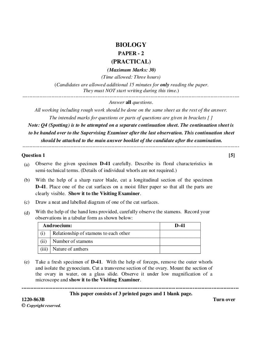
PAPER - 2
(PRACTICAL)
(Maximum Marks: 30)
(Time allowed: Three hours)
(Candidates are allowed additional 15 minutes for only reading the paper.
They must NOT start writing during this time.)
Answer all questions.
All working including rough work should be done on the same sheet as the rest of the answer.
The intended marks for questions or parts of questions are given in brackets [ ]
Note: Q4 (Spotting) is to be attempted on a separate continuation sheet. The continuation sheet is
to be handed over to the Supervising Examiner after the last observation. This continuation sheet
should be attached to the main answer booklet of the candidate after the examination.
Question 1 [5]
(a) Observe the given specimen D-41 carefully. Describe its floral characteristics in
semi-technical terms. (Details of individual whorls are not required.)
(b) With the help of a sharp razor blade, cut a longitudinal section of the specimen
D-41. Place one of the cut surfaces on a moist filter paper so that all the parts are
clearly visible. Show it to the Visiting Examiner.
(c) Draw a neat and labelled diagram of one of the cut surfaces.
(d) With the help of the hand lens provided, carefully observe the stamens. Record your
observations in a tabular form as shown below:
Androecium: D-41
(i) Relationship of stamens to each other
(ii) Number of stamens
(iii) Nature of anthers
(e) Take a fresh specimen of D-41. With the help of forceps, remove the outer whorls
and isolate the gynoecium. Cut a transverse section of the ovary. Mount the section of
the ovary in water, on a glass slide. Observe it under low magnification of a
microscope and show it to the Visiting Examiner.
This paper consists of 3 printed pages and 1 blank page.
© Copyright reserved.
Page 2
(g) Based on your observation in Q.1(e), complete the table given below:
Gynoecium: D-41
(i) Position of the ovary with respect to other floral whorls
(ii) Type of placentation
(iii) Number of locules in the ovary
(h) Write the floral formula of the specimen D-41.
(i) Draw the floral diagram of the specimen D-41.
(j) Name the family to which the specimen D-41 belongs.
(k) Write two characteristic features of the family mentioned by you in Q.1(j) above.
(l) Mention the botanical name (including genus and species) of one economically
important plant belonging to this family.
Question 2 [3]
(a) You are provided with two soil samples D-42 and D-43.
(b) Weigh 10 grams of soil sample D-42 and transfer it into a 100 ml beaker labelled A and
weigh 10 grams of soil sample D-43 and transfer it into a 100 ml beaker labelled B.
(c) Now add 50 ml of distilled water to each of the beakers A and B and stir the contents,
using a glass rod.
(d) Allow the contents to settle down for 10 minutes.
(e) Transfer 10 ml of clear supernatant of the soil sample D-42 from beaker A into a
test tube labelled T1 and transfer 10 ml of supernatant of the soil sample D-43 from
beaker B into a test tube labelled T2.
(f) Place test tubes T1 and T2 in a test tube stand. Determine the pH of the solution in
test tubes T1 and T2 separately with the help of the pH papers provided to you.
Show the pH readings to the Visiting Examiner
(g) Record your results in a tabular form as shown below:
Test tube pH of the supernatant Nature of the soil
(acidic/ alkaline/neutral)
T1
T2
(h) Name one factor that affects the soil pH.
(i) What is the optimum pH of the soil for proper growth of plants?
2
1220-863B
Page 3
(a) With a sharp razor blade, cut several longitudinal sections of the specimen D-44
provided. Select a good section and stain with safranin. Mount it in glycerine on a
glass slide using cover slip.
Show your slide to the Visiting Examiner under a low power Microscope.
(b) Identify the given specimen D-44.
(c) Draw a neat labelled diagram of the mount as seen under the microscope.
(d) Write two characteristic features of this specimen.
Question 4 [5]
Identify the given specimens A to E. Give two reasons to support your answer in each case.
Draw a neat labelled diagram of each specimen. You are not allowed to spend more than
three minutes for each spot.
Note: Hand over your continuation sheets to the Supervising Examiner after you finish
answering this question.
Question 5
Show the following to the Visiting Examiner for assessment:
(a) Project [10]
(b) Biology Practical File. [5]
3
Document Details
| Board / Org | CISCE |
| Exam | Class 12 |
| Type | Question Paper |
| Pages | 3 |
| Language | english |
| Updated | 22 Jul 2026 |