NEET Biology Question Bank - Animal Tissue
Finished viewing? Save it for later —
Download NEET Biology Question Bank - Animal Tissue (PDF · 26 pages)About NEET Biology Question Bank - Animal Tissue
NEET Biology Question Bank - Animal Tissue is available here for free download. Published by NTA for NEET UG, this question bank can be viewed online or downloaded as a PDF (26 pages). Candidates preparing for NEET UG can use NEET Biology Question Bank - Animal Tissue to understand the exam pattern, the type of questions asked, and the overall difficulty level.
Frequently Asked Questions
How can I download NEET Biology Question Bank - Animal Tissue?
Open this page and click the Download button to save NEET Biology Question Bank - Animal Tissue as a PDF. It is completely free on AglaSem Docs.
Is NEET Biology Question Bank - Animal Tissue free to download?
Yes. NEET Biology Question Bank - Animal Tissue can be viewed online and downloaded as a PDF free of cost on AglaSem Docs.
How many pages does NEET Biology Question Bank - Animal Tissue have?
NEET Biology Question Bank - Animal Tissue contains 26 pages, which you can read online or download together as a single PDF.
Where can I find more NEET UG study material?
You can find more NEET UG question papers, sample papers, syllabus, and answer keys on AglaSem Docs.
NEET Biology Question Bank - Animal Tissue – Text
Read the full text of this question bank below — useful to quickly search, copy and reference the content online without downloading the PDF.
📄 View text version (26 pages)
Page 1
· Body Organization : Body organisation process remains dynamic where multicellularity resulted
with complexity. Group of cells structurally and functionally became similar and organised tissue.
Cells ® Tissues ® Organs ® Organ systems ® Complex body.
· There are four basic tissue types :
(1) Epithelial tissue (2) Connective tissue
(3) Muscular tissue (4) Nervous tissue
Epithelial tissue Ñ
Definition : A tissue which covers external surface of the body and interal free surface of many
organs.
Features :
· It forms external surface and internal surface or many organ and body.
· The cells are arranged very close to each other with very little extracellular material or matrix which
is a product of these epithelial cells.
· The cells are thin, polygonal, cuboidal or columnar. They rest on a non-cellular basement
membrane.
Location :
· The lining of the skin, alimentary canal, blood vessels, digestiveglands, respiratory organs, etc.
Function :
· It is highly specialised for protection, absorption and secretion.
· The skin and most of the organs are covered by a epithelial membrane for protection.
· Some of the epithelial surfaces are absorptive and secretory.
Group :
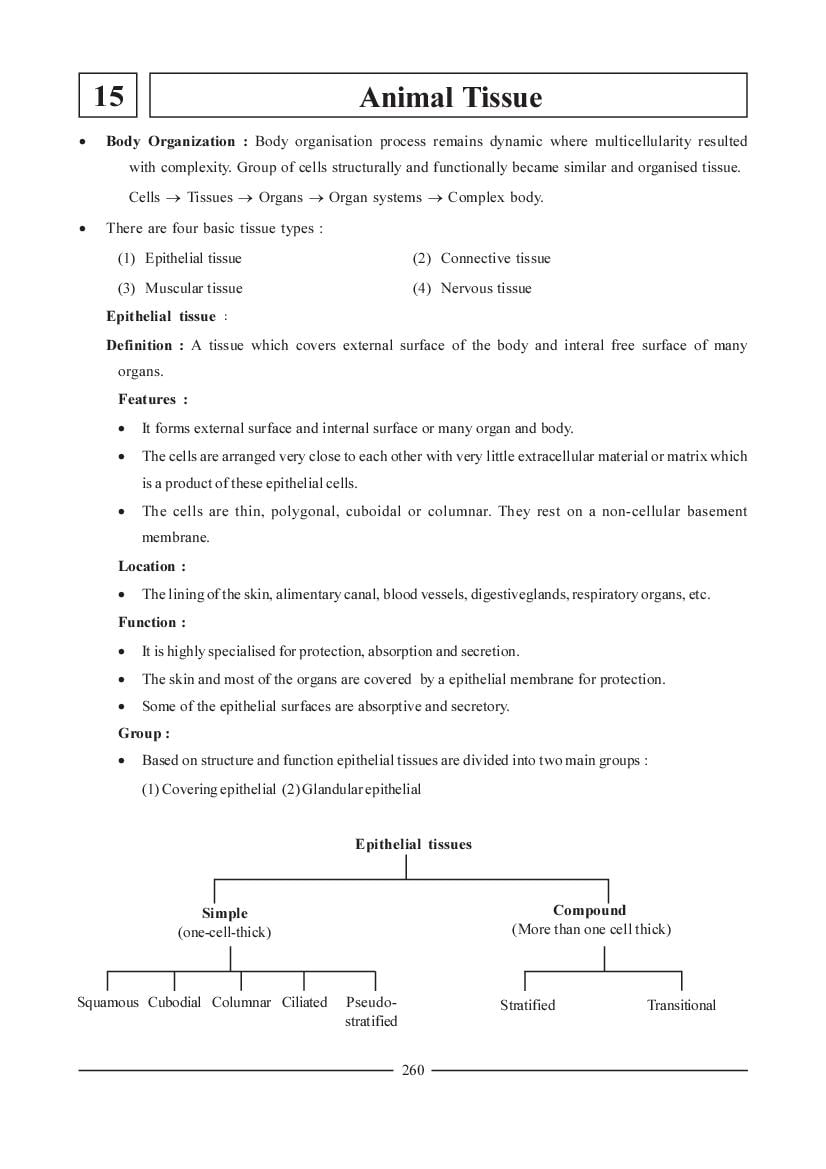
· Based on structure and function epithelial tissues are divided into two main groups :
(1) Covering epithelial (2) Glandular epithelial
Epithelial tissues
Simple Compound
(one-cell-thick) (More than one cell thick)
Squamous Cubodial Columnar Ciliated Pseudo- Stratified Transitional
stratified
260
Page 2
Location :The outer most layer of skin of frog, inner lining of lung alveoli, lining of blood vessels,
Bowman's capsules of kidney, peritoneum of body cavity.
· Function : Protection of underlying tissue.
Features :
· Cells are joined by cement material.
· In surface view, this tissue seems to be composed of flat tiles like that of a pavement.
· Cuboidal epithelial tissue :
Location : Proximal tubules of kidney, salivary glands, pancreatic ducts, thyroid gland and ovary.
Function : In addition to protection, these cells are participate in secretion (gastric juice, hormone,
etc.), excretion and absorption.
Features :
· Cells are square in vertical sections, polygonal in horizontal section.
· Cells of absorptive surfaces often bear microvilli on their free ends.
· Columnar epithelial tissue :
Location : It forms the lining of the mucous membrane of the stomach, gall bladder, urinogenital
organs and their ducts.
Function : Secretion and absorption. Reabsorption and secretion in uriniferous tubules and absorption
of digested food in alimentary canal.
Features :
· Cells of this type of epithelia are elongated and are placed side by side like tall pillars.
· Their inner ends are generally narrow but free ends are broad having polygonal surface.
· Ciliated epithelial tissue :
Location : Auditory canal, uterine tube, respiratory tract.
Function : Cilia moves particle, free cells or mucus in a specific direction.
Feature : It is merely a modification of columnar epithelium.
· Cilia are developed from basal granules of cytoplasm.
· Cells bear thin protoplasmic processes on their surfaces called cilia and hence this tissue is called
ciliated epithelium.
· Pseudo - stratified epithelium tissue :
Location : Inner lining of trachea, large bronchi.
Function : Removes mucus and secretions.
Features : It is the simple columnar epithelium.
· The regular arrangement of the cells is distorted.
· Cells are arranged in single layer, but appears multi-layered, which is a false appearance.
261
Page 3
Definition : "A tissue which has more than one layer". It is divided in two types. (1) Stratified
compound epithelium tissue.
Location : It is present where there is much wear and tear. Such as in the epidermis of skin, the
lining of mouth cavity, the tongue, oesophagus and vagina of mammals.
Function : Protection of underlying tissues.
Types : It has two subtypes :
(1) Stratified cuboidal epithelial, the superficial cells are cuboidal in shape. Such tissue is
present in the inner surface of larger salivary and pancreatic ducts.
stratified squamous non-keratinised and stratified squamous keratinised are two types of
stratified squamous epithelium.
(2) Transitional epithelium tissue : It is present in the passage of the excretory organs.
(1) Epithelial tissue means ......... .
(A) A tissue which forms outer and inner surface of many organs of animal
(B) Unilayered tissue and with intercellular fluid
(C) Many layered tissue and no intercellular fluid
(D) (B) and (C) both
(2) Simple epithelial tissue means ......... .
(A) Many layered tissue, more intracellular fluid
(B) Unilayered tissue, no intracellular fluid
(C) Unilayered tissue, more intracellular fluid
(D) Many layered tissue, no intracellular fluid
(3) Which epithelial tissue forms internal wall of blood vessels ?
(A) Cuboidal (B) Squamous
(C) Stratified (D) Columnar
(4) What is the function of intestinal villi ?
(A) Absorption of food (B) Increases absorptive surface
(C) Controls intestinal movement (D) Prevents movement of food
(5) Which pair is mis-matched ?
(A) Squamous - Frog skin (B) Cuboidal stratified - Oesophagus
(C) Columnar - Peritoneum of frog (D) Ciliated - Bronchi
(6) The nucleus of squamous epithelium is having x and y characteristics.
(A) x = Weak, flat, polygonal, y = Spherical or oval
(B) x = Thin, flat, polygonal, y = Spherical or oval
(C) x = Thin, square, uniangular, y = Hexagonal or oval
(D) x = Thick, flat, polygonal, y = Spherical or oval
(7) x is the tissue whose cells use connection by "Cement".
(A) Cuboidal (B) Columnar (C) Cilicated (D) Squamous
(8) The shape of cuboidal tissue x in Longitudinal section and y in transeverse section.
(A) x = Polygonal, y = Square (B) x = Spherical, y = Oval
(C) x = Square, y = Polygonal (D) x = Polygonal, y = Rod shape
262
Page 4
(A) Cuboidal (B) Squamous (C) Transitional (D) Columnar
(10) Which epithelial tissue is present in skin which joins eyeball and eyelids ?
(A) Cuboidal stratified (B) Stratified epithelial
(C) Columnar epithelial (D) Transitional epithelial
(11) The epithelial tissue which has brush like free surface is found in .........
(A) Testis (B) Stomach (C) Small intestine (D) Ovary
(12) Which epithelial tissue is present in oesophagus, mouth cavity, cornea, vagina and cervix ?
(A) Transitional (B) Columnar
(C) Keratinized stratified (D) Non-keratinized stratified
(13) Whose cells are arranged side by side and like pillar ?
(A) Cuboidal (B) Ciliated
(C) Columnar (D) Squamous
(14) Which group of organs, have cuboidal epithelium ?
(A) Pancreatic ducts, Thyroid glands, Bowman's cup
(B) Proximal tubules of kidney, Salivary glands, Ovary
(C) Pancreatic ducts, Peritoneum of body-coelom, Ovary
(D) Pancreatic ducts, Thyroid glands, Testes
(15) Pseudostratified tissue remains on internal surface of x and y and removes mucus.
(A) x = Trachea, y = Large bronchioles (B) x = Trachea, y = Small bronchioles
(C) x = Pharynx, y = Fine bronchioles (D) x = Oesophagus, y = Small intestine
(16) x tissue removes mucus in particular direction and y tissue removes mucus.
(A) x = Pseudostratified, y = Ciliated epithelium
(B) x = Ciliated epithelium, y = Columnar epithelium
(C) x = Pseudostratified, y = Cuboidal epithelial
(D) x = Ciliated epithelium, y = Pseudostratified
(17) Which tissue is responsible for division and regeneration ?
(A) Epithelium (B) Muscular (C) Connective (D) Nervous
(18) Which tissue forms glands ?
(A) Muscular (B) Epithelium (C) Nervous (D) Connective
(19) Which tissue is present in Reproductive cells ?
(A) Simple epithelial (B) Nervous (C) Connective (D) None of the given
(20) The outer layer of skin is made up of keratin, Because ......... .
(A) It is secretory and protects against wear and tear
(B) It is widely spreaded overall body
(C) It has more thickness
(D) It prevents entry of pathogens
263
Page 5
(A) Joins loosely with each other and forms unevenlayer
(B) Constantly divides to support organs
(C) Becomes rigid and supports organs
(D) Makes unilayer by the help of cement material
(22) Nasal cavity, fine bronchioles, oviductal funnel has tissue ?
(A) Ciliated (B) Columnar (C) Cuboidal (D) Germial epithelial
Answers : (1-A), (2-B), (3-B), (4-B), (5-C), (6-B), (7-D), (8-C), (9-A), (10-B), (11-C), (12-D),
(13-C), (14-B), (15-A), (16-D), (17-A), (18-B), (19-A), (20-A), (21-D), (22-A)
· Connective tissue Ñ
Definition : A tissue which connects together the other tissues of the body.
Features :
· It is the group of cells and matrix containing of intercellular substances secreted by cells themselves.
· Cells are consequently widely spaced.
· They are divisible.
· It forms from mesoderm.
Functions :
· To connect up structures.
· To form packing around organs.
· To replace tissue which have been destroyed by injury.
· To combat foreign toxins.
· To form a supporting framework.
Connective tissue
Connective tissue proper Skeletal tissue Fluid tissue (Blood)
Areolar White Ligament Blood Lymph
tissue fibrous
Bone Cartilage
tissue
Adipose Tendon
tissue
Spongy Compact Hyaline White fibro Yellow elastic
cartilage cartilage
Calcified
cartilage
264
Page 6
(1) Areolar tissue Ñ
· This is the simplest and most widely distributed connective tissue. It is also called loose connective
tissue.
· It is made up of fibres and cells. Fibres are of two types :
· White fibers : It is wavy, unbranched and are arranged in bundles. It is made up of protein collagen.
It can bear weight, pressure or tension.
· Yellow fibers : It is straight, branched and are not arranged in bundle. It is made up of elastin and
is elastic. It forms delicate network. It cannot bear weight, pressure or tension.
· Cells are of three types, i.e. Fibroblasts; Macrophages and Mast cells.
· Mast cells secretes heparin, histamine and serotonin.
(2) Adipose tissue Ñ
Location : Occurs in abundance in the subcuteneous beneanth the skin, around kidney,
mesenteries, bone marrow.
Functions : As it is present as subcutenous tissue, it helps to conserve the body heat.
Structure : Besides Adipocytes. It contains fibroblasts, macrophages, collagen fibers and elastic
fibers.
(3) White Fibrous tissue Ñ
Location : It is mainly present in the Periosteum of the bones and perichondrium of cartilage.
They are also seen at the joint between skull bones and make the immovable.
Function : Connects bones and muscles
· Forms periosteum and perichondrium of bones and cartilage respectively.
· Forms immovable joints (sutures).
Function : White fibers are arranged compactly and parallel in bundles which are made up of collagen
white protein. Fibroblast are present in between white fibers.
(4) Tendon and Ligament Ñ
Tendon :
· It is made up of collagen fibers (matrix).
· Skeletal muscles remain connected with tendon.
Ligament :
· Branched yellow elastin fibers are present.
· The ligament connects the joints and holds them in position.
· Skeletal tissue :
( i ) Cartilage Ñ
· It is partially, strong, soft and elastic.
· Matrix is dense.
· It is special type of skeletal connective tissue, but, it differs from other connective tissue. It has types
as follow.
265
Page 7
(1) Hyaline cartilage Ñ
Location : It is present in larynx, trachea, sternum, hyoid and ribs, etc.
Structure : The word hyaline is derived from the correct word "hylos" which means "glass" and the
hyaline cartilage was so named because in the gross, it appears as a clear bluish coloured
glassy surface.
· Its matrix is clear, homogenous translucent and lacking fibers.
· It is always covered by a tough fibrous membrane.
· It contains blood vessels from which nutritive substances diffuse through the cartilage.
· The cartilage cells are chondroblasts, which secretes chondrin.
· Cells lie in group of two, four or eight in fulfilled space called lacunae.
Function : It protects bones against friction and wear-tear as it has elasticity and limited flexibility.
(2) White fibrous cartilage Ñ
Location : It is typically found in the intervertebral discs, which binds the vertebrae in mammals.
Structure : It consist of dense white fibrous tissue arranged in bundles with cartilage cells between the
bundles.
· The cartilage cells are usually ovoid in shape and are surrounded by matrix.
Function : It protects vertebrae from wear and tear during movement.
(3) Yellow elastic cartilage Ñ
Location : This type of cartilage is found in the pinna, tips of nose, epiglottis, in certain other regions.
Function : It contains yellow elastic fibers.
(4) Calcified cartilage Ñ
Location : It occurs in the pubis of old frogs, in supra scapula and at the head of humerus and femur.
Structure : · It differs from the hyaline type its matrix impregnated with lime salts.
· It is seen in a normal stage in the development of cartilage during early embryonic life but
it is also found in a permanent tissue in the external ears of many mammals.
( ii ) Bone Ñ
Features :
· It is highly vascular.
· It is mineralized.
· It is constantly changing.
· It is hard and rigid.
· It is resilient.
· It has a regenerating capacity.
· It has a canalicular system.
Mature Bone : It is composed of two tissue.
(1) Compact bone and (2) Spongy bone
· The ground substance or the matrix, composed of protein called ossein.
266
Page 8
carbonate, magnesium phosphate and calcium fluorides.
( 1 ) Compact Bone :
· In adult bone flat irregular spaces called lacuna occurs in the solid matrix.
· Each lacuna contains a flat bone cell or osteocyte.
· The lacuna are in communication with one another by fine canalicules.
· In a long dried bone of frog, large number of lamella are present in a ground substance.
· In mammalian bones many column like structures are seen called Haversian system.
· It each Haversian system, several concentric layers (lamellae) of bony matrix encircle a longitudinal
central canal (Haversian canal).
( 2 ) Spongy Bone :
· Spongy bones are found in vertebrae, ribs, skull, etc.
· It contains red bone marrow.
· Red bone marrow is a seat for formation of RBCs (erythrocytes) and granulocytes.
· Liquid connective tissue (Blood) :
( I ) Blood :
· It is an opaque turbid fluid. It is matrix.
· Liquid portion of this tissue is blood plasma.
· Blood cells are of two types : RBCs and WBCs.
· The fragments of cytoplasm that are present in the blood are called platelets.
· Blood differs from other connective tissue in these ways :
(1) Blood cells are different from cells of other connective tissue.
(2) Secretion of matrix is not entirely by blood cells.
(3) Blood cells are not formed by the division of the pre-existing corpuscles.
Blood
Plasma Blood cells Blood platelets
Erythrocytes Leucocytes
Granulocytes Agranulocytes
Neutrophils Eosinophils Basophils Monocytes Lymphocytes
267
Page 9
-
This is the fluid matrix or intercellular substance.
-
55 % amount occurs in blood by plasma.
-
It is almost colourless having faint yellow tinge.
-
It is alkaline and having pH 7.4.
· Blood cells :
· Erythrocytes (Red Blood Corpuscles - RBCs).
· The normal human erythrocyte is biconcave disc.
· The red colour of erythrocyte is due to the presence of haemoglobin, a red coloured pigment which
is a conjugated protein made up of globin and Fe+2 (Ferrous) containing haeme. It has high affinity
for oxygen.
· Shape of RBCs : The shape and size of RBCs vary in different animals. The cells are nucleated in
all vertebrates, except mammals. In mammals it is biconcave.
· No. of RBCs : Under normal condition the blood of adult male contains 41,00,000 to 60,00,000
erythrocytes per cubic milliliter, while that of adult female contains 39,00,000 to 55,00,000 per cubic
milliliter.
· Life span : Life span is about 120 days.
· Origination of RBCs : From Red bone marrow.
· Function : It participates in transporting carbon dioxide from tissue to lungs, as it contains enzyme
carbonic anhydrase.
No. of RBCs Life Span of RBCs
No. Name of Organism No. of RBCs No. Name of Organism No. of RBCs
1. Male (Human) 41,00,000 to 60,00,000 / 1 Cubic milliliter 1. Mammals and 120 days or 4 months
of blood Human
2. Female (Human) 39,00,000 to 55,00,000 / 1 Cubic milliliter 2. Rabbit 80 days
of blood
3. Human Embryo 85 lacs / 1 Cubic milliliter of blood 3. Frog 100 days
4. Rabbit 70 lacs / 1 Cubic milliliter of blood 4. New born baby 100 days
5. Frog 4 lacs / 1Cubic milliliter of blood
· Haemocytometer is used to calculate the Blood cells.
· Leucocytes :
They are small nucleated semi transparent cells devoid of haemoglobin.
· The leucocytes are capable of changing their shape and moving independently through the intercellular
spaces among tissue. They are originated from bone marrow.
· The number of these cells in adult humans are 7.5 ± 3.5 × 10³ per cubic millimeter of blood.
· The number of cells depends upon the condition of the body. During infection of the body they
generally increase in number. They are known as phagocytes because they feed on the bacteria and
broken down tissue cells by engulfing particles.
· Types of WBCs Ñ
· It has two types : Granulocytes and Agranulocytes
· According to chemical nature of granules, granulocytes are of three kinds :
268
Page 10
· The neutrophils whose granules stain weakly with both the acidic and basic stain. They have may
lobed nucleus.
· It is 60 - 70 % of total WBCs.
(ii) Eosinophils (Acidophils) :
· It can be stain by acidic dyes.
· It has bilobed nucleus and cells are large size.
· It is 2 - 4 % of total WBCs.
(iii)Basophils :
· It can be stained by Methylene blue.
· It has "S' shaped nucleus. It is 0.1 to 1 % of total WBCs.
(2) Agranulocytes :
· It is divided in to two types.
· It has medium alkaline cytoplasm with no lobe in nucleus.
(i) Monocytes (ii) Leucocytes
· Largest WBCs · Smallest WBCs
· Nucleus is kidney shape · Large spherical nucleus
· 4 - 8 % of total WBCs · 25 % of total WBCs
· Platelets :
· It is known as thrombocytes also. They are relatively small non-nucleated.
· They are especially concerned with the clotting of blood.
· They are made in the bone marrow.
· General function of blood Ñ
· Transports oxygen. · Transport of food materials.
· Clotting of blood. · To neutralise the toxins.
· Disposal of cell wreckage. · Transport of waste matters.
· Transport and removal of carbon dioxide.
· Transport of hormones, antitoxins, etc.
· Equalization of body temperature.
(23) Which option is correct for given statements for function of connective tissue ?
(1) Removes mucus.
(2) Connects structures.
(3) Combat foreign toxins.
(4) Participates in excretion and absorption.
(5) To form a supporting frame work.
(6) To replace tissue which have been destroyed by injury.
(A) 1, 2 Right Statements; 3, 4, 5 and 6 Wrong Statements.
(B) 1 and 4 Wrong Statements; 2, 3, 5 and 6 Right Statements.
(C) 2, 3 and 4 Right Statements; 1, 5 and 6 Wrong Statements.
(D) 3, 4 and 5 Right Statements; 1, 2 and 6 Wrong Statements.
269
Page 11
(A) White fibrous (B) Adipose
(C) Areolar (D) Hyaline cartilage
(25) White fibres are x and yellow fibers are y in Areolar tissue.
(A) x = Wavy and branched, y = Thin, less, not in bundle and in bundles
(B) x = Wavy, unbranched, not in bundle.
y = Less, Thin and in bundle.
(C) x = Wavy, unbranched, in bundle.
y = Less, thin, not in bundle.
(D) x = Wavy, branched, in bundle.
y = More, thick and not in bundle.
(26) Which protein is present in white fibers and yellow fibers of Areolar tissue respectively ?
(A) Elastin, Collagen (B) Collagen, Keratin
(C) Collagen, Fibrinogen (D) Collagen, Elastin
(27) Which one is wrong statement ?
(A) Mast cells and Basophils secretes histamine and heparin.
(B) Life is long for mast cell, while it is short for Basophils.
(C) Mast cells are smaller, Basophils are binucleated.
(D) Mast cells are stable while basophils are movable.
(28) Which cartilage has a major role in recovery of nasal septum of Rudransh if it has been injured
during cycling ?
(A) Compact cartilage (B) Calcified cartilage (C) Hyaline cartilage (D) Spongy cartilage
(29) Which tissue combat against the toxins which have been entered into the body of Rudransh ?
(A) Epithelial (B) Connective (C) Skeletal muscle (D) Nervous
(30) Which option is proper for secretion of collagen ?
(A) Histocytes (B) Fibroblast (C) Mast cells (D) Phagocytes
(31) Which pair is true for location of Hyaline cartilage ?
(A) Larynx, Trachea, Sternum, Hyoid, Ribs
(B) Larynx, Bonemarrow, Mesenteries, Sternum, Ribs
(C) Larynx, Subcutaneous, Around Kidney, Sternum, Ribs
(D) Larynx, Trachea, Sternum, Hyoid, Bone marrow
(32) Which tissue provides materials for the attachment of the muscles ?
(A) Epithelial (B) Skeletal (C) Nervous (D) Muscular
(33) The intervertebral discs are made up of .....
(A) Elastic cartilage (B) Fibrous cartilage (C) Calcified cartilage (D) Hyaline cartilage
270
Page 12
(A) White fibrous cartilage (B) Calcified cartilage
(C) Hyaline cartilage (D) Yellow elastic cartilage
(35) Which tissue is present on the head of bone of Rana tigrina ?
(A) White fibrous (B) Calcified cartilage
(C) Hyaline cartilage (D) Yellow elastic cartilage
(36) Which option is correct for location of spongy bone ?
(A) Scapula, Ribs, Skull (B) Ileum, Ribs, Skull
(C) Vertebrae, Ribs, Skull (D) Patalla, Vertebrae, Skull
(37) Which option is proper to maintain cells in bone marrow ?
(A) Osteocytes (B) Chondrocytes (C) Osteoclast (D) Bidder canal
(38) What is called a joint between two Haversian canals ?
(A) Semi circular canal (B) Volkman's canal (C) Inguinal canal (D) Bidder canal
(39) Which structure is jointed by Volkman's canal ?
(A) Matrix - Haversian canal (B) Two Haversian canals
(C) Two different bones (D) Matrix - Osteocytes
(40) Which inorganic salts are present in ossein of skeletal matrix ?
(A) Calcium Chloride, Calcium Carbonate, Magnesium Phosphate
(B) Calcium Phosphate, Calcium Carbonate, Magnesium Phosphate, Calcium Flooride
(C) Pottassium Chloride, Magnesium Phosphate, Calcium Carbonate, Calcium Phosphate
(D) None of these
(41) Which option is correct for frog's bone marrow ?
(A) Yellow, Adipose tissue, Blood vessels (B) Green, Adipose tissue, Blood vessels
(C) Yellow, Hyaline cartilage, Blood vessels (D) Red, Adipose tissue, Blood vessels
(42) Which compound of blood is protective ?
(A) Albumin (B) Agglutinin
(C) Hormones (D) Salts of Pottassium
(43) Which one is non-protein group in the respiratory pigment of human RBCs ?
(A) Mg+2 (B) Fe+2 (C) Ca+2 (D) Zn+2
(44) Which are active phagocytes in WBCs ?
(A) Neutrophils, Monocytes (B) Eosinophils, Lymphocytes
(C) Neutrophils, Eosinophils (D) Lymphocytes, Basophils
(45) To which function the globulin of human blood plasma will be responsible ?
(A) Immunity (B) Maintain osmotic pressure
(C) Conduction of oxygen (D) Blood clotting
(46) During resting condition, the process of formation of RBCs will be ..........
(A) Increased (B) Decreased
(C) Unchanged (D) None of the given
271
Page 13
(A) Basophils > Eosinophils > Neutrophils
(B) Eosinophils > Basophils > Neutrophils
(C) Neutrophils > Eosinophils > Basophils
(D) Eosinophils > Neutrophils > Basophils
(48) Which is the correct quantity of Haemoglobin in 100 ml of Human blood ?
(A) 511 gram (B) 2530 gram
(C) 1720 gram (D) 12160 gram
(49) In the structure of Haemoglobin .............
(A) 70 % globin + 30 % Haematin (B) 80 % globin + 20 % Haematin
(C) 95 % globin + 05 % Haematin (D) 90 % globin + 10 % Haematin
(50) What is the proportion of RBCs and WBCs in blood ?
(A) 1 : 1000 (B) 1000 : 1 (C) 5 : 3000 (D) 3000 : 5
(51) Which blood cells can change their shape ?
(A) WBCs (B) RBCs (C) Platelets (D) All of the given
(52) Which is the waste material in blood ?
(A) Immunoglobulin (B) Thrombin (C) Fibrinogen (D) Carbon dioxide
Answers : (23-B), (24-C), (25-C), (26-D), (27-C), (28-A), (29-B), (30-B), (31-A), (32-B), (33-B),
(34-D), (35-B), (36-C), (37-A), (38-B), (39-B), (40-B), (41-A), (42-B), (43-B), (44-A), (45-A),
(46-B), (47-A), (48-D), (49-C), (50-C), (51-A), (52-D)
Muscular Tissue
· Muscular tissue :
(Muscle tissue)
· It is formd from mesoderm. It consists of cellular
elements in the form of fibres of various length. Skeletal muscle Simple muscle Cardiac
muscle
· No intercellular liquid is present. or or
· All muscular tissue have great contractility. Striated muscle Non striated muscle
or or
· By contraction and relaxation, it make possible
Voluntary muscle Involuntary
the movement of whole body.
or
· It has three types. Smooth muscle
· Skeletal muscle or striated muscle :
· The muscle fibers are the thin units of skeletal muscle. Each muscle fibre is a single thin and
elongated cell provided with many nusclei.
· The muscle fibers are arranged in bundles (fasciculi).
· In higher animals, they are found associated with the skeleton.
· They are attached to bones by tendons.
· Muscle fibres can be contracted as desired. So, they are also known as voluntary muscles.
· Because of their appearance they are called striated or striped muscle.
· Structure of muscle fibers Ñ
· The cytoplasm (sarcoplasm) of each fiber contains large number of myofibrils.
· Each fiber has an extensive sheath, the sarcolemma.
272
Page 14
· The darker bands are called "A" band.
· The lighter bands are called "I" band.
· Each I band is divided by middle dark line, is known as z-disc or Krause's membrane.
· Two successive z-disc covers the region - "Sarcomere".
Myofibrils
Thick Thin
Made up of Myosin Made up of Actin,
Tropomyosin, troponin protein
· Each sarcomere consists of one full A-band and two halves I-bands.
· In middle of A-band, the actin fibres are absent. So, it looks something lighter. It is known as
H-band or Hensen's zone.
· Smooth muscle or Involuntary muscle :
Location : This type of tissue is present in visceral organs - like oesophagus, stomach, intestine, blood
vessels and pupil, etc.
Structure :
· It is made up of mononucleated spindle shape cells.
· It has granular Sarcoplasm around its nucleus. These muscles always receive their nerve supply
from autonomous nervous system.
· Cardiac Muscle :
Location : In the wall of Heart.
Structure :
· It appears as network of branching and anastomosing cylinders.
· The space between the cardiac muscle cells are occupied by the endomysium. The endomysium
contains fibroblasts. Collagen and reticular fibres.
· Each cell of cardiac muscle shows striation with A, I, Z and H-bands.
· At the end of the cardiac muscle cells, there are prominent cross striation called intercalated discs.
· Nucleus is arranged deep into sarcoplasm between two successive intercalated discs.
· These muscles are capable of contracting rhythmically and are immune to fatigue. There is rich
blood supply.
· It is innervated by autonomous nervous system.
(53) What is the location of H-band ?
(A) Middle portion of myosin fibers of A-band
(B) Middle portion of Actin fibers in A-band
(C) Middle portion of myosin fibers of I-hand
(D) Middle portion of Actin fibers in Z-band
273
Page 15
(A) x = Mechanical (B) x = Heat (C) x = Electric (D) x = Light
(55) What are the symbols for dark band, Krause's membrane, Hensen's zone, light band ?
(A) A, C, H, I (B) I, Z, H, A (C) A, Z, H, I (D) I, C, H, A
(56) What is structural unit for muscle ?
(A) Myosin (B) Actin (C) Sarcomere (D) Tropomyosin
(57) Actin and myosin is connected to .......
+ +
(A) Na and K pump (B) Muscle contraction
(C) Nervous system (D) Absorption of water
(58) Where is the strongest muscles are present in Human body ?
(A) Jaw (B) Thigh (C) Neck (D) Hands
(59) Where is the location of biggest muscle in Human ?
(A) Chick (B) Neck (C) Ear (D) Hip muscle
(60) In straited muscle, the thick and thin myofibrils are made up of proteins known as...
(A) Myosin, Collagen (B) Myosin, Troponin
(C) Myosin, Actin-Tropomyosin-Troponin (D) Keratin, Actin-Myosin-Troponin
(61) Which option is correct for smooth muscle ?
(A) Cylindrical, unbranched, straited, multinucleated and voluntary
(B) Spindle, unbranched, non-straited, mononucleated, involuntary
(C) Cylindrical, unbranched, straited, multinucleated, involuntary
(D) Spindle, unbranched, non-straited, multinucleated, involuntary
(62) What is the type of contraction of cardiac muscle ?
(A) Rhythmic, immune to fatigue (B) Rhythmic, not immune to fatigue
(C) Rapidly, not immune to fatigue (D) Rapidly, immune to fatigue
(63) How cardiac muscle defers from Non-straited muscle ?
(1) Intercalated disc (2) Autonomous (3) About nucleus
(A) 1 and 2 (B) 1 (C) 1, 2 and 3 (D) 2 and 3
(64) What is the name of dark and light band respectively ?
(A) I, A (B) A, I
(C) H, Z (D) Z, H
(65) What is Sarcomere ?
(A) Region between two successive Z-band (B) Region between two I-band
(C) Region between two successive A-band (D) The surface of two Z-bands
Answers : (53-B), (54-A), (55-C), (56-C), (57-B), (58-A), (59-D), (60-C), (61-B), (62-A), (63-C),
(64-B), (65-A)
274
Page 16
Nervous tissue
Neurons Neuroglia
- Long processes - Short processes
- It conducts nerve impulse - It supports neurons
· Structure of Neurons :
· Typically each neuron consist of a cell body and branching fibres.
· Cell-body : It is called cyton, cytoplasm of cyton contains a large and spherical nucleus. There are
large number of dark particles called Nissl's granules.
Processes (or fibres)
(Fibres arises from periphery of cyton)
Axons Dendrites
- It is longer - It is shorter
- Conducts impulse away from neuron - Conducts impulse towards the neuron
- It is called efferent - It is called afferent
Types of Neurons
Unipolar Bipolar Multipolar
- Only one process - One axon and one - More than two processes
- e.g. In Embryo dendrite - e.g. Brain, Spinal Cord
- e.g. Retina
· The nerve fibres may be surrouned by two concentric sheaths. The inner is known as medulla or
myelin sheath.
· It is surrounded by a transparent cellular sheath, the neurilemma.
· The neurilemma sheath is made up of single layer of flat expanded Schwann's cells.
· Each myelinated nerve fiber shows constrictions at regular intervals called nodes of Ranvier.
· The nerve endings of axon are not in direct physical contact with nerve endings of dendrites of
another neuron. This physical gap is called synapse.
· Nerve impulses pass between neurons through the synapse with the help of a hormone Acetylecholine
called Neurotransmitters.
(66) Which fibre conducts the impulse towards cyton ?
(A) Axon (B) Dendrite (C) White fibres (D) Yellow fibres
275
Page 17
(A) Gastrin (B) Glucagon (C) Collagen (D) Acetylcholine
(68) What is the composition of Nissl's granules ?
(A) Mitochondria (B) Golgi body (C) Lysosome (D) Ribosome
(69) The myelin sheath is present in which tissue ?
(A) Epithelial (B) Connective (C) Muscular (D) Nervous
(70) Which type of Neurons are present in the mammalian brain ?
(A) Monopolar (B) Multipolar (C) Bipolar (D) All of the given
(71) Which type of neuron is present in mammalian retina of eyes ?
(A) Monopolar (B) Multipolar (C) Bipolar (D) All of the given
(72) Which type of neuron is present in Embryo ?
(A) Monopolar (B) Multipolar (C) Bipolar (D) All of the given
Answers : (66-B), (67-D), (68-D), (69-D), (70-B), (71-C), (72-A)
· True and Flase (T - F) Type Questions :
(73) Which option is correct for given statement ?
(1) The cells of squamous epithelium are thick, flat and polygonal.
(2) The cells of cuboidal epithelium in longitudinal section are polygonal.
(3) The internal end of the cells of columnar epithelium are broad and narrow at free end.
(4) Ciliated epithelium is modified columnar epithelium.
(A) F, T, F, T (B) T, T, F, F (C) F, T, T, F (D) F, F, T, T
(74) (1) Pseudostratified epithelium is simple columnar epithelium.
(2) Pseudostratified epithelium removes mucus in particular direction.
(3) Transitional epithelium remains in organ of excretory system.
(4) Stratified epithelium provides durable covering to the organs.
(A) F, F, T, T (B) T, F, T, T (C) T, T, F, F (D) T, F, T, F
(75) In reference to connective tissue which option is correct ?
(1) It connects the organs.
(2) Combat foreign toxins.
(3) Makes suture in skull bones.
(4) Adipose tissue remain beneath the skin.
(A) T, T, T, T (B) T, F, T, T (C) F, T, T, T (D) F, F, T, T
(76) (1) Simplest and widely spreaded tissue is areolar.
(2) White fibrous tissue is in tendon and is inelastic.
(3) White fibers are made up of elastic protein and yellow fibres are of collagen protein.
(4) Blood is opaque and turbid liquid.
(A) T, T, F, F (B) F, F, T, T (C) T, T, F, T (D) T, F, T, F
276
Page 18
(1) Conducts oxygen (2) Do not conduct waste
(3) Neutralises toxins (4) Do not maintain temperature
(A) T, T, F, F (B) F, F, T, T (C) F, T, F, T (D) T, F, T, F
(78) Which option is correct for skeletal muscle ?
(1) A-band dark and has myosin
(2) I-band is light and has actin
(2) A-band contracts during muscle contraction
(4) Sarcomere is a region between two successive Z-band
(A) T, T, F, T (B) T, T, F, F (C) F, F, T, T (D) T, T, T, T
(79) Which option is correct in reference to Nervous tissue ?
(1) Neurons have long processes which conducts impulses.
(2) Neuroglia has short processes and provide support to Nervous tissue.
(3) Monopolar Neuron has only one process.
(4) Bipolar neuron has one axon and one dendrite.
(A) T, F, T, F (B) T, T, F, F (C) T, T, T, T (D) F, F, F, F
Answers : (73-A), (74-B), (75-A), (76-C), (77-D), (78-A), (79-C)
(80) Match the column :
Column - I Column - II
(1) Squamous epithelium (A) Intestinal glands (A) 1 - A, 2 - B, 3 - D, 4 - C, 5 - E
(2) Cuboidal epithelium (B) Trachea (B) 1 - E, 2 - D, 3 - B, 4 - A, 5 - C
(3) Columnar epithelium (C) Ovary (C) 1 - D, 2 - E, 3 - A, 4 - B, 5 - C
(4) Ciliated epithelium (D) Blood vessels (D) 1 - D, 2 - C, 3 - A, 4 - E, 5 - B
(5) Pseudostratified (E) Uriniferous tubules
(81) Match the column :
Column - I Column - II
(1) Squamous epithelium (A) Square in longitudinal section (A) 1 - A, 2 - C, 3 - B, 4 - D
(2) Ciliated epithelium (B) Very thin and flat cells (B) 1 - D, 2 - B, 3 - C, 4 - A
(3) Cuboidal epithelium (C) Polygonal cells and broad at free end (C) 1 - B, 2 - D, 3 - A, 4 - C
(4) Columnar epithelium (D) Modified columnar epithelial cells (D) 1 - C, 2 - A, 3 - D, 4 - B
(82) Match the column :
Column - I Column - II
(1) Epithelial tissue (A) Combat foreign toxins (A) 1 - A, 2 - D, 3 - C, 4 - B
(2) Connective tissue (B) Capacity for contraction (B) 1 - B, 2 - A, 3 - C, 4 - D
(3) Muscular tissue (C) Conduction of nerve impulse (C) 1 - A, 2 - B, 3 - C, 4 - D
(4) Nervous tissue (D) Arrange on basal membrane (D) 1 - D, 2 - A, 3 - B, 4 - C
277
Page 19
Column - I Column - II
(1) Neutrophils (A) Spherical large nucleus (A) 1 - D, 2 - E, 3 - B, 4 - A, 5 - C
(2) Eosinophils (B) S shaped nucleus (B) 1 - A, 2 - C, 3 - D, 4 - B, 5 - E
(3) Basophils (C) Kidney shaped nucleus (C) 1 - C, 2 - B, 3 - A, 4 - E, 5 - D
(4) Lymphocytes (D) 2-7 lobed nucleus (D) 1 - B, 2 - A, 3 - C, 4 - D, 5 - E
(5) Monocytes (E) Bilobed nucleus
(84) Match the column :
Column - I Column - II
(1) Squamous epithelium (A) Beneath the skin (A) 1 - D, 2 - A, 3 - C, 4 - B
(2) Hyaline cartilage (B) Bowman's capsule (B) 1 - B, 2 - D, 3 - A, 4 - C
(3) Adipose tissue (C) Wall of stomach (C) 1 - A, 2 - B, 3 - D, 4 - C
(4) Simple muscle (D) Hyoid bone (D) 1 - D, 2 - C, 3 - B, 4 - A
(85) Match the column :
Column - I Column - II
(1) Tendon (A) Matrix (A) 1 - C, 2 - D, 3 - A, 4 - B
(2) Ligament (B) Ossein (B) 1 - A, 2 - D, 3 - C, 4 - B
(3) Hyaline cartilage (C) Collagen fibers (C) 1 - B, 2 - D, 3 - C, 4 - A
(4) Bone (D) Chondrine (D) 1 - C, 2 - A, 3 - D, 4 - B
Answers : (80-D), (81-C), (82-D), (83-A), (84-B), (85-D)
· A - Assertion and R - Reason type questions :
Which option is correct for given question ?
(A) A and R both correct, R is correct explanation of A.
(B) A and R both correct, R is not correct explanation of A.
(C) A - correct, R - wrong.
(D) A - wrong, R - correct.
(86) Assertion A : Squamous epithelium is like mosaic tiles at a surface view.
Reason R : Cells of this tissue is joined by cement material.
(A) (B) (C) (D)
(87) Assertion A : The cells of squamous epithelia are thin and flat.
Reason R : Main function of this tissue is of protection.
(A) (B) (C) (D)
(88) Assertion A : The cells of cuboidal tissue are polygonal in L.S. and square in T.S.
Reason R : Its function is protection, secretion, excretion and absorption.
(A) (B) (C) (D)
278
Page 20
Reason R : Function of this tissue is secretion and absorption.
(A) (B) (C) (D)
(90) Assertion A : Ciliated tissue is modified into columnar tissue.
Reason R : Pseudostratified epitheliam is a kind of simple epithelium tissue.
(A) (B) (C) (D)
(91) Assertion A : White fibres are wavy and branched in Areolar tissue.
Reason R : White fibers are made up of collagen protein.
(A) (B) (C) (D)
(92) Assertion A : Adipose tissue helps in maintaining body temperature.
Reason R : Suture of skull is occupied by white fibrous tissue.
(A) (B) (C) (D)
(93) Assertion A : Cartilage differs from normal connective tissue.
Reason R : It contains dense matrix.
(A) (B) (C) (D)
(94) Assertion A : In bones of mammals, they have large amount of tubular structures in it. It is
called haversian system.
Reason R : It is seen in sponge bone, vertebrae, ribs and skull.
(A) (B) (C) (D)
(95) Assertion A : Serum is part of blood other than required components for blood clotting.
Reason R : Human RBCs are biconcave discoid shape.
(A) (B) (C) (D)
(96) Assertion A : Nucleus of Monocyte is kidney shaped.
Reason R : Monocytes are smallest WBCs.
(A) (B) (C) (D)
(97) Assertion A : Platelets are also known as Thrombocytes.
Reason R : The secretes Thromboplastin.
(A) (B) (C) (D)
(98) Assertion A : WBCs are called as phagocytes.
Reason R : They engulf bacteria and broken cells.
(A) (B) (C) (D)
(99) Assertion A : Skeletal muscle is also known as voluntary muscle.
Reason R : They can contract according to desire of an animal.
(A) (B) (C) (D)
279
Page 21
Reason R : It is seen in alimentary canal and pupil.
(A) (B) (C) (D)
(101) Assertion A : Cardiac muscle has cross striation at end, is called intercalated discs.
Reason R : Comparision to Z-disc, it is thinner.
(A) (B) (C) (D)
(102) Assertion A : Blood differes from other connective tissue.
Reason R : Blood cells do not secretes matrix.
(A) (B) (C) (D)
(103) Assertion A : Dendrites are afferent in nature.
Reason R : They conduct impulse towards the cyton.
(A) (B) (C) (D)
(104) Assertion A : Axon are efferent in nature.
Reason R : They conduct impulse away from cyton.
(A) (B) (C) (D)
(105) Assertion A : Monopolar Neuron has only one process.
Reason R : In this Neuron dendrite and axon are same fibers.
(A) (B) (C) (D)
(106) Assertion A : Monopolar Neurons are present in Retina.
Reason R : Monopolar neurons has one fibers only on one side.
(A) (B) (C) (D)
(107) Assertion A : Multipolar neurons has more than two processes.
Reason R : Bipolar neurons has two processes.
(A) (B) (C) (D)
(108) Assertion A : Sarcoplasm of neuron has large and oval shape nucleus.
Reason R : Cyton has some dark granules, which are called as Nissl's granules.
(A) (B) (C) (D)
(109) Assertion A : Axon and dendrite are not in direct physical contact.
Reason R : Acetylcholine act as a neurotransmitters.
(A) (B) (C) (D)
Answers : (86-B), (87-C), (88-D), (89-B), (90-D), (91-D), (92-B), (93-A), (94-B), (95-B), (96-C),
(97-A), (98-A), (99-A), (100-B), (101-C), (102-A), (103-A), (104-A), (105-A), (106-D), (107-B),
(108-D), (109-B)
280
Page 22
(110) Which tissue is mentioned in given figure ? e b
(A) Columnar (B) Ciliated c
(C) Pseudostratified (D) Cuboidal d
(111) What is indicated by a, b and c in given figure ?
(A) a = Basal granules, b = pili, c = Nucleus (B) a = Nucleus, b = pili, c = Basal granules
(C) a = Basal membrane, b = pili, c = Nucleus (D) a = Cilia, b = Basal granules, c = Nucleus
(112) What is indicated by d and e in given figure ?
(A) d = Basement membrane, e = mucus secreting cells
(B) d = Nucleus, e = Basal granules
(C) d = Mucus secreting cells, e = Basal membrane
(D) d = pili, e = Mucus secreting cells a
b
(113) Which tissue is mentioned in given figure ?
c
(A) Columnar (B) Ciliated
d
(C) Pseudostratified (D) Cuboidal
f e
(114) What is indicated by a, b and c in given figure ?
(A) a = Nucleus, b = pili, c = Basal granules (B) a = Cilia, b = Basal granules, c = Nucleus
(C) a = Basal membrane, b = pili, c = Nucleus (D) a = Basal membrane, b = pili, c = Nucleus
(115) What is indicated by d, e and f in given figure ?
(A) d = Basal cell, e = Mucus secreting cell, f = Basal membrane
(B) d = Basal membrane, e = Basal cell, f = Mucus secreting cell
(C) d = Basal cell, e = Basal membrane, f = Mucus secreting cell
(D) d = pili, e = Basal cell, f = Mucus secreting cell a
(116) Which tissue is indicated in given figure ?
b
(A) Pseudostratified (B) Ciliated
(C) Cuboidal (D) Stratified epithelium c
d
(117) Match the Column by help of the given figure :
Column - A Column - B
(p) Basement membrane (i) b (A) (p-ii), (q-i), (r-iii), (s-iv)
(q) Epithelial layer (ii) d (B) (p-ii), (q-i), (r-iv), (s-iii)
(r) Germinative layer (iii) a (C) (p-ii), (q-iv), (r-i), (s-iii)
(s) Dead cells (iv) c (D) (p-ii), (q-iii), (r-i), (s-iv)
281
Page 23
a
(A) a = Ligament, b = Muscle, c = Bone, d = Tendon
b
(B) a = Ligament, b = Bone, c = Muscle, d = Tendon
c (C) a = Ligament, b = Bone, c = Tendon, d = Muscle
d
(D) a = Tendon, b = Muscle, c = Muscle, d = Ligament
(119) Which tissue is indicated by given figure ?
(A) White elastic cells (B) Yellow elastic cell
(C) Hyaline cartilage (D) White fibrous tissue
(120) What is indicated by a, b, c and d in the given figure ?
(A) a = Cartilage cell, b = Matrix, c = Collagen fiber, d = Lacuna d
a
(B) a = Collagen fibers, b = Lacuna, c = Matrix, d = Cartilage cells
(C) a = Collagen fibers, b = Matrix, c = Lacuna, d = Chondroblast
(D) a = Matrix, b = Chondroblast, c = Lacuna, d = Collagen fibers b c
(121) What is indicated by a, b and c in given figure ?
(A) a = Haversian system, b = Interstitial lamellae,
a b
c = Concentric lamellae
c
(B) a = Concentric lamellae, b = Haversian system,
c = Interstitial system d
(C) a = Interstitial lamellae, b = Haversian system,
e
c = Concentric lamellae
f
(D) a = Haversian lamellae, b = Canaliculi,
c = Haversian system g
(122) What is indicated by d, e, f and g in given figure ?
(A) d = Lacunae with bone cell, e = Matrix, f = Haversian system, g = Canaliculi
(B) d = Concentric lamellae, e = Matrix, f = Haversian system, g = Canaliculi
(C) d = Haversian system, e = Canaliculi, f = Matrix, g = Haversian tubules (canal)
(D) d = Canaliculi, e = Matrix, f = Haversian system, g = Haversian tubules (canal)
(123) Identify the parts of striated muscle in given figure ?
(A) a = Sarcoplasm, b = Nucleus, c = Sarcolemma,
d = Muscle fibre, e = Dark band, f = Light band f
(B) a = Sarcoplasm, b = Light band, c = Muscle fibers, a
d = Sarcolemma, e = Nucleus, f = Dark band
(C) a = Light band, b = Sarcoplasm, c = Muscle fibers, b
c
e
d = Sarcolemma, e = Nucleus, f = Dark band
(D) a = Sarcolemma, b = Nucleus, c = Dark band,
d
d = Light band, e = Sarcoplasm, f = Muscle fibers
282
Page 24
a
Column - A Column - B b
(p) Nucleus (i) h
c
(q) Dendrite (ii) i
(r) Node of Ranveir (iii) b i d
(s) Nerve fibers (iv) c e
(t) Schwann's cell (v) d f
j
(u) Nerve fibers (axis) (vi) e g
(A) (p-iv), (q-iii), (r-v), (s-i), (t-ii), (u-vi)
h
(B) (p-iv), (q-iii), (r-v), (s-ii), (t-i), (u-vi)
(C) (p-iv), (q-ii), (r-iii), (s-v), (t-i), (u-vi)
(D) (p-iv), (q-vi), (r-ii), (s-iii), (t-i), (u-v)
(125) What is indicated by a, f, g and j by given figure ?
(A) a = dendrite, f = Myelin sheath, g = Nerve fiber, j = Nucleus
(B) a = Nissl's granule, f = Myelin sheath, g = Nerve fiber, j = Nucleus
(C) a = Nissl's granule, f = Nerve fiber, g = Myelin sheath, j = Nucleus
(D) a = Nissl's granule, f = Nucleus, g = Myelin sheath, j = Nerve fiber
Answers : (110-B), (111-D), (112-A), (113-C), (114-B), (115-A), (116-D), (117-B), (118-A), (119-A),
(120-B), (121-C), (122-A), (123-C), (124-A), (125-B)
· Questions for NEET :
(126) Blood vessels are made up of which cells ?
(A) Columnar epithelial (B) Connective tissue
(C) Simple muscle tissue (D) Squamous epithelium
(127) Where is the location of columnar tissue in Human ?
(A) Intestine and Urethra (B) Auditory canal and Wall of stomach
(C) Bronchus and Fallopian tube (D) Stomach and Oesophagus
(128) Which tissue is present in layer of Alveoli ?
(A) Simple epithelium (B) Cuboidal epithelium
(C) Squamous epithelium (D) Ciliated epithelium
(129) Collagen is made up of ......... .
(A) Lipid (B) Carbohydrate (C) Globular protein (D) Fibrous protein
(130) Ligaments means
(A) Modification of white elastic fibers (B) Modification of yellow elastic fibers
(C) Inelastic white fibers (D) None of the given
(131) Areolar tissue connects ......... .
(A) Bone to bone (B) Lipid to muscle
(C) All the muscles (D) Muscle to bone
283
Page 25
(A) Hyleuronic acid (B) Basement membrane
(C) Collagen fibers (D) Liquid
(133) Which fibers of striated muscles are connected with the activity of ATPase ?
(A) Actin (B) Troponin (C) Tropomyosin (D) Myosin
(134) What is called the cell of Bone ?
(A) Chondroclast (B) Osteoclast (C) Lecuna (D) Osteocytes
(135) To whom the globulin protein of human blood plasma is connected ?
(A) Immunity (B) Maintenance of osmotic pressure
(C) Conduction of oxygen (D) Blood clotting
(136) Which WBCs has highest amount in blood ?
(A) Neutrophils (B) Eosinophils (C) Basophils (D) Lymphocytes
(137) WBCs are true cells, Because ......... .
(A) Nucleus is present (B) Acts as a phagocytes
(C) It is polymorphism (D) None of the given
(138) How much Haemoglobin is present in human RBCs ?
(A) 40 % of biomass (B) 34 % of biomass
(C) 90 % of biomass (D) 50 % of biomass
(139) Which option is correct for content of Nissl's granules ?
(A) Cellular molecular (B) Lipid molecules (C) Ribosomes (D) Mitochondria
(140) What is called the connection of axon of a neuron with dendrite of another neuron ?
(A) Constant bridge (B) Synapse (C) Joint (D) None of the given
Answers : (126-D), (127-C), (128-C), (129-D), (130-B), (131-C), (132-B), (133-D), (134-D),
(135-A), (136-A), (137-A), (138-B), (139-C), (140-B)
284
Page 26
¯
¯ ¯ ¯ ¯
Epithelial tissues Connective tissues Muscular Nervous tissues
tissues - Controls the body
- One or more layers. - Structural frame, support to - Contractive according to stimulation
body. and body response.
- Outer and inner layer - Becomes
of organ or body. - Interstitial liquid, cells and active by - It has conduction,
electricity co-ordination and
fibers.
- Helpful in response.
movement - Nerve cells, Neuroglial
® Simple Epithelium cells, Neuroganglion,
etc.
® Semiliquid/Semi solid
® Squamous mosaic
files like flat and ® Areolar tissue ® Skeletal muscle
single layer, polygo- Connects different Elongated and unbranched
nal cells body parts muscles. Joined with bones.
® Cuboidal Small, ® Adipose tissue
cube, shapecells, Villi Eye ball and around
® Smooth muscle
at free end of cells, kidney Elongated and spindle shape fibers.
Brush like Villi Involuntary muscles. Visceral muscles.
for absorption
® Columnar Elongated ® Tendor ® Cardiac muscle
cells like pillar Joins muscle to bone
Elongated and cylindrical fibers.
® Ciliated Diverse in ® Ligament Never get fatigue. Antonomous.
shape with nucleus Joins bone to bone
with cilia at free end
Cartilage tissue
®
Semi-solid matrix,
® Supportive Hyaline, Fibrous, Caclified
skeletal tissue and Elastic
Compound
® Epithelium Bones
® Keratinised, ® Hardest tissue, Solid matrix,
non keratinised, Bony and spongy bones
Transitional tissue is
with 4 - 6 layers ® Blood
® Transitional epithelial ® Liquid Red liquid, RBCs,
tissue : tissue WBCs platelets
- 4 to 6 layers
- maintains thickness
It shows
Lymph
re-organisation ® Pale yellow, Blood plasma and
Glandular Epithelium WBCs
®
Secretory in nature. In glands
·
285
Document Details
| Board / Org | NTA |
| Exam | NEET UG |
| Type | Question Bank |
| Pages | 26 |
| Updated | 22 Jul 2026 |