GSEB HSC Model Question Paper for Biology - Set 2
Finished viewing? Save it for later —
Download GSEB HSC Model Question Paper for Biology - Set 2 (PDF · 22 pages)About GSEB HSC Model Question Paper for Biology - Set 2
GSEB HSC Model Question Paper for Biology - Set 2 is available here for free download. Published by Gujarat Board for Class 12, this sample paper can be viewed online or downloaded as a PDF (22 pages). Candidates preparing for Class 12 can use GSEB HSC Model Question Paper for Biology - Set 2 to understand the exam pattern, the type of questions asked, and the overall difficulty level.
Frequently Asked Questions
How can I download GSEB HSC Model Question Paper for Biology - Set 2?
Open this page and click the Download button to save GSEB HSC Model Question Paper for Biology - Set 2 as a PDF. It is completely free on AglaSem Docs.
Is GSEB HSC Model Question Paper for Biology - Set 2 free to download?
Yes. GSEB HSC Model Question Paper for Biology - Set 2 can be viewed online and downloaded as a PDF free of cost on AglaSem Docs.
How many pages does GSEB HSC Model Question Paper for Biology - Set 2 have?
GSEB HSC Model Question Paper for Biology - Set 2 contains 22 pages, which you can read online or download together as a single PDF.
Where can I find more Class 12 study material?
You can find more Class 12 question papers, sample papers, syllabus, and answer keys on AglaSem Docs.
GSEB HSC Model Question Paper for Biology - Set 2 – Text
Read the full text of this sample paper below — useful to quickly search, copy and reference the content online without downloading the PDF.
📄 View text version (22 pages)
Page 1
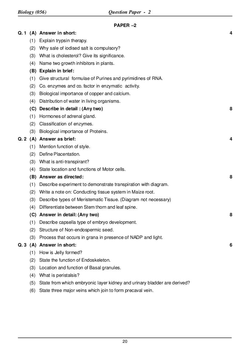
PAPER –2
Q. 1 (A) Answer in short: 4
(1) Explain trypsin therapy.
(2) Why sale of iodised salt is compulsory?
(3) What is cholesterol? Give its significance.
(4) Name two growth inhibitors in plants.
(B) Explain in brief:
(1) Give structural formulae of Purines and pyrimidines of RNA.
(2) Co. enzymes and co. factor in enzymatic activity.
(3) Biological importance of copper and calcium.
(4) Distribution of water in living organisms.
(C) Describe in detail : (Any two) 8
(1) Hormones of adrenal gland.
(2) Classification of enzymes.
(3) Biological importance of Proteins.
Q. 2 (A) Answer as brief: 4
(1) Mention function of style.
(2) Define Placentation.
(3) What is anti-transpirant?
(4) State location and functions of Motor cells.
(B) Answer as directed: 8
(1) Describe experiment to demonstrate transpiration with diagram.
(2) Write a note on: Conducting tissue system in Maize root.
(3) Describe types of Meristematic Tissue. (Diagram not necessary)
(4) Differentiate between Stem thorn and leaf spine.
(C) Answer in detail: (Any two) 8
(1) Describe capsella type of embryo development.
(2) Structure of Non-endospermic seed.
(3) Process that occurs in grana in presence of NADP and light.
Q. 3 (A) Answer in short: 6
(1) How is Jelly formed?
(2) State the function of Endoskeleton.
(3) Location and function of Basal granules.
(4) What is peristalsis?
(5) State from which embryonic layer kidney and urinary bladder are derived?
(6) State three major veins which join to form precaval vein.
20
Page 2
(B) Answer the following questions: 6
(1) Describe : Cardiac muscular tissue.
(2) Write a note on Autonomous Nervous system.
(3) Functions of liver with respect to excretion.
(C) Describe in detail: (Any two) 8
(1) Organogenesis from mesoderm
(2) Excretory system of frog.
(3) Internal structure of skin and its function.
Q. 4 (A) Answer briefly : 4
(1) What are Contained genes ?
(2) Why petite mutant yeast produces fewer ATP moleucles ?
(3) What is Philadelphia syndrome ?
(4) What are okazaki fragments ?
(B) Complete linkage rarely occurs – Explain through Morgan’s experiment on Drosophiila. 3
(C) Answer as directed: (Any two) 8
(1) Explain in detail operon model gene regulation.
(2) Explain the phenomenon of sex determination in man.
(3) Inheritance of Kappa particler in Paramecuim.
(D) Draw labeled diagram: (Any one) 5
(1) Pectoral girdle.
(2) Open Buccopharyngeal cavity of frog.
Q. 5 (A) Answer briefly: 6
(1) What is anaphylactic shock?
(2) Explain green house effect.
(3) State full form of ELISA and POM.
(4) Name the carcinogens causing cancer of liver.
(5) What is social forestry?
(6) What is the contribution of Cohen?
(B) Answer as directed: (Any two) 8
(1) Role of micro-organisms in nitrogen cycle.
(2) Symptoms of hepatitis.
(3) Functions of health centers.
(4) Write a note of tissue culture technique.
(C) Answer any two : 6
(1) What are B cells? How do they react against an antigen?
(2) Cancer therapy.
(3) Write a Note on AIDS.
21
Page 3
ANSWER
A. 1 (A) Answer in short.
(1) Explain trypsin therapy.
Trypsin is an enzyme secreted by the pancreas, which in pure form, is given to the patient to cure
ultcres. This is known as tryspin therapy.
(2) Why sale of iodised salt is compulsory?
Iodine is an essential constituent of thyroxius – a thyroid hormone secreted by the thyroid gland.
Its deficiency affects the physical, mental and reproductive development. Hence Government
insists on the sale of iodised salt.
(3) What is cholestrol? Give its significance.
Cholestrol is a sterol (an important type of lipid.) It is a common structural material of the nervous
tissue. It is also present in meet, liver animal fat. If its amount increases in the blood, the walls of
the blood vessels become stiff and the flow of the blood through the vessels is impaired.
(4) Name any two growth inhibitors in plants.
(i) Ethylene (ii) Abscissin
A. 1 (B) Explain in brief.
(1) Purines and pyrimidines of RNA.
RNA has two purines Adenine and Guanine, and two pyrimidines cytosine and uracil. Their
structure are as follows:
(2) Coenzymes and co-factors are the prosthetic groups or the non-protein part of an enzymes.
Some enzymes become active only in the presence of a prosthetic group. Coenzymes are organic
compounds. Which act as hydrogen – acceptors and get reduce while participating in the enzymatic
reaction.
Examples of coenzymes include:
NAD – Nicotinamide adenine dinucleotide
NADP – Nicotinamide adenine dinucleotide phosphate
FMN – Flavin mononucleotide
FAD – Flovin adenine dinucloetide
Co-factors : Co-factors are mineral ions which activate the enzymes. e.g.s. of co-factor are
22
Page 4
magnesium, calcium, Zinc manganese copper etc. Enzymes enolase is activated only in the
presence of Mg++, Manganese and Zinc.
(3) Biological infoilance of copper and calcium.
Copper: is important in the synthesis of haemoglobin and chlorophyl. Enzymes like tyrosienase
have copper as a structural component. Copper is present in haemocyanin a respiratory pigment
in certain arthropods.
Calcium: In animals, calcium in the main component of bones and teeth.
It is an indispensible element for (1) blood co. agulation (2) maintainance of PH,
(3) exlitation of nervers and muscles and (4) development of bones.
In mants it playp an unetectaut take in.
(1) Structural architescture of cell wall
(2) Permeability of cell membrane.
(3) Converting organic toxins to non toxic forms.
(4) Distribution of water in living organisms.
In living organisms, water is distributed as (1) extracellular water and (2) intracellular water.
Water in the blood plasma, intercellular fluid, cerebro-spinal fluid, optic fluid and lymph is
extracellular water. Intracellular water molecules are in two forms : Bound form and free
form. Water molecules surrounding certain protein or other large molecules or their groups
are in the bound form. Water has the property of polarity due to which molecules or their
groups present in solution are arranged in a specific order, which forms the bound form of
water. Certain ions are firmly bound to water molecules while others are loosely bound.
The protoplasm is the main structural support of the cells of living organisms. In fact,
chemically it is most complex. This form is either positively or negatively charged. Hence
water molecules are arranged in specific manner only. Due to their polarity, the ions present
in water get arranged in two layers.
Arrangement of water molecules in the complex
(C) Describe in details: (any two)
(1) Hormone of adrenal gland
The adrenal gland is imbedded in the tissue of the kidneys in frog. It has two parts (1) the medulla
and (2) the cortex. The medulla of adrenal gland secrete adrenaline and non adrenaline.
Adrenaline regulater the heart beats, constriction – dilation of blood vessels and blood
supply to voluntary muscles, It also includes conversion of glycogen to glucose, increases
rate of respiration, retards peristatic movement of stomach and intestine wall. It also enhance
the production of ACTH in the anterior pituitary.
Nor- adrenaline regulates the constriction – dilation of blood vessels to maintain blood
pressure.
23
Page 5
The cortex of the adrenal gland secretes Aldosterone corticosterone and
hydrocorticsosterone.
Aldosterons influence the rate of reabsorption of minerals and water in the urini ferous
tubules. Its secretion is not influence by the level of ACTH.
Corticosterone and hydrocortisone affect the induction of proteins and liphid metabolism
throgh carbohydrate metabolism. Their secretion is regulated by ACTH.
Deficiency of of cortilosteoids causes loss of sodium and water in the body resulting in
fatigue feeling of thirst and lack of maintenance of blood glucose level.
(2) Classification of enzymes :
According to international union of Biochemistry enzymes are classified into six groups these are.
(1) Oxidoreductases (2) transferases (3) Hydrolases (4) lyases (5) Isomerasers (6) ligases
or synthetases
(1) Oxidoreductases: Thisgroup of enzymes are associated with Oxidation-reduction reaction in
the cell. The enzymes which remove hydrogene form the substrate are known as dehydrogenase
and the ones which adds oxygen are known as oxidases eg. Succinic dehydrogenase, oytochromo
oxidse of ETS.
(2) Transferases : These enzymes bring about the transfer of a group form one substrate to another
substrate. Eg. Hexokinase which transter phosphate group of ATP to an hexose sugar in the first
reaction of glycolysis.
ATP ADP
Glucose → glucose – 6 – phosphate
Hexokinase
(3) Hydrolases : Split complex organic substance by adding a molecule of water
Maltose + H2o
→ glucose + glucose
Maltase
(4) Lyases : Cleave macromolecules without adding water eg aldolase
aldolase
Fructose – 1, 6 diphosphate → DHAP + PGAL
(5) Isomrases : Catalyze the changes in the molculal structure of the substrate of glucose – 6 –
phosphate isomease
→ fructose 6 – phosphate
(6) Ligase : There enzymes synthesize a new moleenle by joining two mokcules with the help of
ATP or acetyl co. A synthtase.
acetic acid + ATP + LO A ← acetyl co. A + AMP + PP.
(3) Biological Importance of Proteins :
(1) Proteins are the main structural components of the membranes of cell organelles.
(2) They are the chief constituents of protoplasm, and they also occur in extracellular fluids.
(3) All enzymes and certain animal hormones are proteins. The hormones of pancreas, pituitary
and parathyroid glands are peptides in natures.
(4) Haemoglobin, is important for the exchange of O2 and Co2.
(5) Immunoglobin in blood plasma has the property of immunity.
(6) Rhodopsin, essential for vision, is a protein.
(7) The contractile proteins like (i) actin and myosin in muscles and globular protein in cilia
and flagella are reponsible for movements.
(8) Melanin, a colour giving pigment, is a protein.
(9) Fibrinogen and thrombin help in the coagulation of blood.
24
Page 6
(10) The egg protein and casein in mammalian milk is used for the development of embryo.
(11) Any two individulas do not resemble completely in their protein structure. Hence variations
are seen in allliving organisms.
A. 2 (A) Answer in Brief :
(1) Mention function of style :
It is a solid, delicate, filamentous structure connecting stigma with ovary.
It arranges stigma in such a way as to enable it to receive the pollen grains and forms a passage
for the pollen tube.
(2) Define : Placentation.
The arrangement of placentae and the ovules in relation to the ovary in called placentation.
(3) What is antitranspirant ?
Liquid wax of low density is sprayed on the plants to minimize the rate of transpiration without
affecting the process of photosynthesis. Such a chemical substance is described as anti
transpirant.
(4) State location and function of Motor cell.
Location: They are located in the upper epidermis of maize leaves in group of 2-5 cells having a
small, unicellular curved trichome on each lateral side.
Function: These cells loose water in dry atmosphere and decrease the leaf surface area by
causing the coiling of leaf margins and thus regulate the rate of transpiration.
(B) Answer As Directed.
(1) Describe experiment to demonstrate transpiration with diagram.
Pieces if filter paper soaked in 5% cobalt chloride solution become dark blue when dried,
when this paper comes in contact with water or vapour it turns pink.
Two pieces of COCl2 paper of equal size
are kept on both the surfaces of a leaf of a potted
plant and then are covered by glass plates and
fixed together by clips or rubber bands. Then the
edges of the glass plates should be covered by a
ccat of Vaseline.
After a few hours it is observed that the
Cocl2 paper on the lower surface of leaf turns pink
more rapidly. It can be inferred from this
observation that the rate of transpiration is higher
from the lower surface as compored to that from
the upper surface of leaf because of large number
of stomata on the lower surface.
This phenomenon can also be Diag Experiment to demonstrate
demonstrated by the 4 – leaf experiment. transpiration.
(2) Write a note on Conducting tissue system in Maize root.
It is located in the stele and possess xylem and phloem bundles. In maize root nearly 20
xylem and an equal number of phloem bundles exist. These bundles of xylem and phloem are
located below the pericyele and are alternatively arranged in the peripheral region of the stele.
Such On arrangement is known as alternate and radial arrangement.
25
Page 7
Since there are several xylem and phloem bundles in stele the stele is described as polyarch
stele.
Each xylem bundles consist one metaxylem vessel towards medulla and two protoxylem
vessels towards the periphery of stele. The metaxylem is surrounded by a layer of
sclernchymatous cells.
Since the protoxylem is towards periphery and metaxylem towards the inner side, the
xylem is described as exarch xylem. Phloem possess companion cells, sieve tubes and phloem
parenchyma in maize root.
(3) Describe types of Meristematic Tissue. (No diagram)
On the basis of position in the plant body meristems are classified into 3 types namely,
(A) Apical meristems,
(B) Intercalary meristems
(C) Lateral meristems
(A) APICAL MERISTEMS :
– It is located at the stem tip, root tip, branch tip, which are the growing points of the plant
body.
– Apical meristem result in growth and elongation of stem, root and branches.
– The apical and axillary buds of stem shows three regions in their longitudinal section.
– They are an outer most unilayered epiblem or dermatogen, a central multilayered plerome
and a multilayerd periblem between these two the apical region of stem also possess a
pair of prophylls.
– The apical region of root apex shows epiblem, periblem and plerome. Apart from this, root
apex shows calyptrogen a multilayered meristematic tissue, which gives rise to root cap.
(B) INTERCALARY MERISTEMS :
– These are the meristems present between the permanent tissue.
– These are actually the portious of apical meristems which gets separated from the apex
during the growth of the axis, and get arranged between permanent tissues.
– This tissue is short lived shows meristematic property for a short time and merge with
permanent tissue, after getting differentiated.
– This tissue are seen in internodes, sheathing leafbases of grasses and in equisetum, a
pteridophyte.
– It result in rapid elongation of internodal portion of the shoot (or) stem.
(C) LATERAL MERISTEM :
– This type of merstem is found on the lateral sides of stem and root.
– It is responsible for the formation of secondary tissues.
– Its activity result in secondary growth due to which organs like stem and roots increases
in diameter.
– Primary intravascular cambium, secondary intervascular cambium and cork cambium
of flowering plant are example of lateral meristems.
26
Page 8
(4) Differentiate between : stem thorn & leaf spine.
Stem thorn Leaf Spine
(1) Modification of axillary or apical bud of (1) Modification of stipules of leaf into a
the stem into a sharp structure for sharp structure is called leaf spine. They
protection is called stem thorn. They may never possess flowers and leaves.
possess flowers and leaves.
(2) A leaf or bud is not present in its axil. (2) A leaf or bud is present in its axil.
(3) Stem thorns are branched (carissa) or (3) Leaf spines are branched.
unbranched (pomegranate).
(4) Stem thorns are endogenous in origin. (4) Leaf spines are exogenous in origin
e.g Thorns of Carissa & Pomegranate. e.g. Zizyphus, Acacia
(C) Answer In Detail :
(1) Describe capsella type of embryo development.
– The development of the embryo and the
endosperm occurs simultaneously.
– The type of development described herein is
capsella type of embryo development.
– In this type of development the zygote divides
transversely into two unequal cells.
– The larger cell towards the micropyle is called
basal cell, while the other smaller cell towards
the center of embryo sac is called apical cell.
– The basal cell divides transversely once again
into two unequal cells, while apical cell divides
longitudinally into two apical cells. These four cells
Diag Development of embryo
are arranged in ^ shape together form a
proembryo.
– In the proembryo the largest and lowermost cell is called a basal call which does not divide any
more. Above the basal cell there is a single middle cell above which are two apical cells.
– The middle cell of the proembryo repeatedly divides transversely and produces a 15 to 20 celled
long filamentous structure called suspensor. Due to the formation of suspensor the apical cells
are pushed towards the endosperm so that the nutritional need of the developing embryo is
satisfied.
– The lowermost cell of the suspensor, towards the micropylar end is the largest and called basal
cell which anchors the proembryo with the wall of embryo sac at the micropylar end.
– The two apical cells first divide longitudinally and then transversely into eight cells forming an
octant ( a group of 8 cells).
– The terminal cell of the suspensor which is closely associated beneath the octant is called
hypophysis cell.
– The cells of the octant divide peripherally to form 16 – celled embryo. Its upper mass of eight
cells is called a terminal or chalagap octant, while the lower mass of eight cells is called a
micropylar octant.
27
Page 9
– The terminal octant develops into two cotyledons and the plumule, while the micropylar
octant gives rise to the hypocotyl and the stepar region (plerome) of the radicle.
– The hypophysis cell divides repeatedly and the cells so formed give rise to the epiblem
(dermatogen) and periblem as well as calyptrogen and and the root cap regions of the radicle.
– The suspensor degenerates when the embryo development is completed.
(2) structure of Non- endopermic seed.
– Bean seed is flat, kidney shaped structure with a notch on one side.
– Along the notch a long white scar exists, in which hilum is present.
– Seed remains attached to the inner margin of the fruit with the hilum.
– A small pore known as micropyle is located at one end of the hilum. Through micropyle only the
water enters into bean seed when it is placed in moist soil.
– Structurally bean seed possess the following part :-
Seed
Seed Coats Embryo
Testa Tegmen Cotyledons Radicle Plumule
SEED COATS:
– Two seed coats exist around bean seed. The outer seed coat is known as testa, it is thick
leathery and relatively strong. The inner seed coat is known as tegmen, it is thin and membranous
both the testa and tegmen are more ot less united.
EMBRYO:
– It is the juvenile form of the plant that remain protected in seed.
– Embryo consists two cotyledons & embryo – axis.
– Embryo- axis possess plumule, hypocotyl & radicle.
– Embryo- axis remain enclosed between two cotyledons in bean seed.
(a) Plumule:
– It is the first embryonic apical bud having a growing point covered by delicate under developed
leaves. Plmule is connected to radicle through hypocotyl. During seed germination plumule
and hypocotyl gives rise to shoot system.
(b) Radicle:
– It is a small, rod shaped part of the embryo axis, it is located towards micropyle region of the
seed. During germination, radicle emerge out first through the micropyle and develops primary
root, which in turn gradually establishes tap root system.
COTYLEDONS:
– Bean seed possess two cotyledons. These structures are fleshy due to stored food in them.
Protein and starch grains exists as stored food. In bean seed, during embryonic development
28
Page 10
cotyledons enclose nutritive endosperm tissue, therefore separate endosperm does not exist in
bean seed. Such seeds are therefore known as exalbuminous or non – endospermic seeds.
Since cotyledons nourish the embryo during seed germination they are known as nurse leaves.
(3) Process that occurs in grana in presence of NADP and light.
Presence of light is necessary for light reaction. It occurs in the grana of Chloroplasts. In this
process release of O2, reduction of NADP and synthesis of ATP (photophosphorylation) occurs.
– Robert Hill proved that photolysis of water occurs with the help of photons of sunlight and the H +
ions released in this process reduce the hydrogen acceptor such as NADP.
– The oxygen molecules released in this process are not released from CO but only from H O.
2 2
This was shown experimentally using the radioactive isotope O18.
– Light reaction occurs in two phase as follows :
(1) Cyclic photophosphorylation.
(2) Non – cyclic photophosphorylation.
– Photophosphorylation is the process of synthesis of ATP molecules by the combination of ADP
with inorganic phosphate by the indirect utilization of solar energy.
(I) CYCLIC PHOTOPHOSPHORYLATION :
– In this process only PS-I participates.
– PS-I (P700 ) is stimulated by absorbing 4 photons from sunlight. As a result 4 energy rich electrons
(4e) are set free from PS-I.
– The 4e liberated from PS-I sequentially pass through different electron carriers viz. unknown
electron carrier x, Ferredoxin reducing substance (FRS), Ferredoxin (FD), Cytochrome – b6,
cytochrome – f and platocyanin (PC) respectively and finally return to PS-I.
– During this course of electron transport one ATP molecule is fomed when elections pass from Fd
to cytochrome – b6 and another ATP is formed when electrons pass from cytochrome b6, to
cytochrome. The energy required for synthesis of ATP are synthesized during cyclic
photophosphorylation.
During this process photolysis of water, reduction of NADP and evolution of O2 does not occur.
(ii) NON – CYCLIC PHOTOPHOSPHORYLATION:-
– Both PS-I and PS-II participate in this process.
– In this process, 4 photons of sunlight stimulate PS-II (P680). As a result four energy rich electrons
(4e) are released from PS-II. These electrons enter into PS-I instead of returning to PS-II.
– Moreover, the 4e released by the stimulation of PS-I neither go back to PS-I nor to PS-II, but are
utilized for the reduction of NADP.
– During non – cyclic photophosphorylation the process such as photolysis of water release of O2,
reduction of NADP and synthesis of one molecule of ATP occur. These can be shown as under:
(1) Under the influence of 4 photons of sunlight the PS-I becomes stimulated and release 4e. These
electrons sequentially pass through various electrons carriers known as x, FRS and FD. Finally
the 4e liberated from FD join with NADP & activate it.
(2) Simulaneously 4 photons of sunlight also stimulate PS-II.AS a result 4e from PS-II are released
which sequentially pass through various electron carriers known as guinone (Q), unknown electron
carrier y, plastoquinone (Pq), cytochrome – f and plastocyanin (PC) and finally enter into PS-I.
(3) Photolysis of water with help of unknown enzymes and in the presence of Mn+2 and Ct, 4 molecules
of water gets dissociated into 4H+ and 40H–.
29
Page 11
enzyme
4H2O → 4H+ + 4 OH−
+2
Mn , ct
(4) The 4OH– so released in the above process form 2H2O, 4e– and a molecule of O2 The O2 molecule
is released into the atmosphere. But the 4e– pass through an unknown electron carrier Z and
then enter the PS-II.
40H − → 2H2 O + O 2 + 4 e −
4 e − → Z → PS − II
(5) The 4H+ ions released as a result of photolysis of water join with activated 2 NADP molecules
with the help of enzyme NADP reductase and reduce it to 2 NADPH2.
NADP
2NADP + 4 e − + 4H+ → 2NADPH 2
reductase
(6) When 4e– pass from electron carrier Pq to cytochrome – f. they lose some energy which is used
to synthesise one molecule of ATP.
A. 3 (A) ANSWER IN SHORT :
(1) How is Jelly formed ?
The eggs when pass through oviduct are enveloped by liquid, viscous albumin which swells by
absorbing water, when comes in contact with it, and becomes very thick transparent and colorless
jelly. This Jelly does not permit the entry of any sperm through it.
(2) State the function of endoskeleton.
The functions of endoskeleton are as follows :
(1) It gives shape to and maintains the shape of a body.
(2) It provides support and protection to the soft parts.
(3) It provides surface for the attachment of muscles
(4) It acts as a lever system.
(3) Location and function of Basal granules.
Location – They located in the cytoplasm of ciliated epithelium.
Function – They give rise to cilia.
(4) What is peristalsis?
The passage of food through the alimentary canal or digestive system due to the rhythmic
30
Page 12
contraction and relaxation of the muscles of the wall of alimentary canal is called peristalsis.
(5) State from which embryonic layer kidney & urinary bladder are derived?
Kidney is derived from mesoderm and urinary bladder is derived from endoderm.
(6) State three major veins which join to form precaval vein.
External jugular, innominate and subclavian vein.
(B) ANSWER THE FOLLOWING QUESTION:
(1) Describe : Cardiac muscular tissue
This is a special type os muscular tissue
that forms the wall of the heart. It is also an
involuntary muscular tissues. Its contraction
and relaxation occurs rhythmically and
never get fatigued. Its fibres, like that in
striated muscles, show A and I bands but
the sarcolemma is relatively less developed
and the nucleus is embedded in the Cardiac muscular tissue
cytoplasm as in non-striated muscle fibres. As special features the cardiac muscles are not as
separate fibres but form a continuous reticulate structure because of the oblique cytoplasmic
brdiges connecting the adjacent fibres at several places. In addition, the fibres show thick and
dark transverse bands, called intercalated discs at regular intervals. Each fibre contains several
nuclei but a single nucleus between two consecutive intercalated discs. These muscles are
innervated by nerve fibres from the vagus nerve and also from autonomous nervous system.
(2) Write a note on Autonomous Nervous System.
The autonomous nervous system comprises of
(a) Sympathetic Nervous System &
(b) Parasympathetic Nervous System.
(a) Sympathetic Nervous System:-
– It is formed of sympathetic ganglia, sympathetic cords and the non-medullated nerve fibres arising
from the sympathetic ganglia.
– A pair of sympathetic chains run along the outer sides of & parallel to the systemic arch &
dorsal aorta.
– Anteriorly these chains enter the cranium & join with the gasserian ganglia. Each gasserian
ganglion is also connected with the vagus ganglion.
– The sympathetic nervous system forms a cardiac plexus around the heart and the associated
parts anteriorly & forms a solar plexus posteriorly.
(b) Parasympathetic Nervous System:-
– It is divided into (1) a Cranial section & (2) a Sacral section.
– Very fine nerve fibres arising from some of the cranial nerves form a nerve plexus which constitutes
the cranial section.
– While the fine nerves fibres arising from the posterior spinal nerves form a nerve plexus that
constitutes the sacral section.
31
Page 13
– The sympathetic and the parasympathetic nervous systems are functionally complementary &
contradictory to each other. e.g The rate of heart beats increases when the parasympathetic
nervous system is excited while it decreases when the sympathetic nervous system is excited.
(3) Functions of liver with respect to excretion.
Excretory Function:
– The toxic ammonia produced due to protein digestion when brought by the blood into liver is
converted in harmless urea.
– The NH2 produced due to deamination of surplus amino acids in liver, is converted by the liver
into harmless urea.
– Liver facilitates disintegration of haemoglobin released into the blood due to haemolysis of RBC
and converts it into bile pigments known as bilirubin & biliverdin which will be excreted out of the
liver a longwith the bile juice.
– Liver excretes calcium salts and cholestrol along with the bile juice.
– Indole, Cresole, Sketole & carbolic acid are the toxic gaseous substances produced due to
incomplete oxidation of proteinous faecal matter by the bacteria in the rectum of digestive system.
When these toxic wastes are brought by blood from rectum into liver they are decomposed to
form harmless urea.
– Liver also disintegrates old damaged R.B.C.’s.
(C) DESCRIBE IN DETAIL:
(1) Organogenesis from mesoderm :
The mesodermal cells lying above the archenteron and beneath the neural plate get differentiated
into vacuolated cells that form a compact, elastic rod-like structure called notochord. The
formulation of the notochord is called notogenesis. The notochord, now, becomes distinctly
distinguishable from the rest of the mesoderm.
The mesoderm that lies on both the lateral sides of the notochord forms a thick, solid layer
which gradually encloses the entire archenteron. Two longitudinal groves develop along each of
the lateral sides that divide the mesoderm into an epimere a mesomere and a hypomere. The
epimere lies dorsally close to the notochord and divides into a few solid, more or less cuboidal
Differentiation in mesoderm
mesodermal somites, arranged in a linear row on each side of the notochord. Such an arrangement
of somites is called metameric segmentation. Gradually each somite gets differentiated into an
outermost dermatome, a myotome and the innermost sclerotome from which the dermis of the
skin, muscles of the dorsal body wall and the skull as well as vertebral column develop
respectively. The mesomere is also termed as nephrotome from which the reproductive organs
32
Page 14
as well as excretory organs, other than urinary bladder, develop. The hypomere is, initially, a solid
band of mesoderm. In the hypomere. On each side, there develops a longitudinal slit that splits
the hypomere into two layers, the outer known as somatic mesoderm and the inner, splanchnic
mesoderm. The space between these two layers gradually enlarges and forms the body cavity
(coelom). The dermis of the skin of the ventral and lateral body wall, the muscels of the body wall,
the appendicular skeletal tissue and other connective tissues develop from the somatic mesoderm,
while the muscles and connective tissues of the alimentary canal develop from the splanchnic
mesoderm. The somatic mesoderm joins with the ectodern and forms somatopleure, while the
splanchnic mesodern joins with the endoderm and forms splanchnopleure. The mesoderm on the
ventral side of the archenteron gives rise to the enitire circulatory system.
(2) Excretory system of frog.
– The organs engaged in the removal
of nitrogenous wastes like urea, uric
acid. Ammonia, creatinine, creatine,
etc. from the body, together form the
excretory system.
– The excretory system of frog
possessing the following structures:
® Excretory organs → Kidneys.
® Ureters
Excretory system ® Urinary bladder
®
® Accessory excretory organ ® ® Cloaca & Cloacal aperture
KIDNEYS:
– Eaca kidney is dorsovertrally flattened & oblong in shape.
– They are dark red in colour.
– The outer margin or surface is simple & slightly convex while the inner margin possesses several
notches.
– An yellow streak like adrenal gland is located on the mild-ventral surface of kidney which runs
longitudinally.
– Kidneys are located in the subvertebral lymph sinus dorsally to, & outside the body cavity.
– The ventral surface of each Kidney is covered by peritoneum.
– Funnel shaped ciliated bodies known as nephrostomes are located on the ventral side of the
kidneys.
33
Page 15
– Nephrostomes are wide externally & narrow internally.
– The broad end opens into body cavity & the narrow end opens into renal reins,
URETERS:
– They arise from the anterior end of kidney & run inside the kidney along the outer margin and
energe posteriorly.
– They move dowmwards and finally open into cloaca from its dorsal side.
URINARY BLADDER:
– It is thin walled, bilobed sac-like structure possessing non – striated muscles in its wall.
– It is located at the ventral wall of the cloaca.
– It opens into ventral wall of cloaca through a large aperture.
– Urinary bladder store urine for expulsion.
CLOACA:
– It is the hindermost part of the alimentary canal.
– The ureters, urinary bladder & in female frog the oviducts open into it.
(3) INTERNAL STRUCTURE OF SKIN & ITS FUNCTIONS.
– The skin of frog is thin, moist, slimy, highly vascularised and devoid of any type of exoskeleton.
– Histologicaley it can be divided into two regions:-
(1) The Epidermis & (2) The Dermis
® Stratified epithelium
® Epidermis←
® Germinative stratum
Skin
® Loose Connective tissue
® Dermis ←
® Compact Connective tissue.
EPIDERMIS :
– It is formed of stratified epithelium which arises from the ectoderm.
– It forms the outer compactly cellular region of the skin.
– The innermost layer of cells of the epidermis rests on basement membrane and consists of –
more or less colummar cells having large distinct nuclei.
– The cells of this layer being capable of dividing the layer is called germinative stratum or
Malphigian layer.
– Its cells divide & produce new cells which do not divide but are pushed continuously toward the
outer side and therefore become more & more flat with diminishing activity.
– The outer layer of cells form the stratified squamous epithelium which is protective in function.
– The outermost layer is removed by friction & replaced by the new cells from within.
– The surface of the epidermis possesses a large number of pores – the openings of the mucous
glands.
DERMIS:
– It is mesodermal in origin.
– It chiefly consists of connective tissue.
34
Page 16
– The dermis consists of an outer, relatively thin layer of loose connective tissue and an inner
thicker layer of compact connective tissue containing tissue containing transverse & longitudinal
strands of muscles, blood vessels & nerve fibres.
– A large number of flask – shaped mucous glands arise from the epidermis.
– The bulbous part of these glands extends in the loose connective tissue of dermis while the
narrow neck remains in the epidermis and opens on the outer surface by a pore.
– The nerve fibres have their nerve endings in the loose connective tissue forming touch
corpuscles.
– The blood vessels capillariees profusely in the loose connective tissue close to the epidermis.
– There are several chromatophores containing melanin pigments beneath the epidermis especially
in the skin of the dorsal & dorsolateral sides of frog.
– The chromatophones impart colour to the skin of frog.
FUNCTIONS OF SKIN :
(1) It forms a complete envelope around the body & renders protection to the internal soft parts of the body.
(2) The slimy covering over the
epidermis protects the body
against bacteria & fungi & helps
to escape easily from the grip
of enemies.
(3) The skin can change its colour
to some extent in order to
match the surroundings and
protect the animal form the
enemies.
(4) The frog does not drink water
but absorbs it through the skin.
(5) The skin acts as a general sense organ.
(6) It is a chief respiratory organ when the frog is on land & the sole respiratory organ when the
animal is in water & in soil.
(7) The skin of ready to hatch tadpole secretes an enzymes that soften and partly dissolves egg
membrane to facilitate hatching.
A. 4 (A) Answer briefly.
(1) Contained genes :
When a single gene segment has other genes in it, such genes are called contained genes. Such
genes are observed in PHI( φ )X-174 virus.
(2) Why petite mutant yeast produces fewer ATP molecules.
Petite mutant yeast have abnormal mitochondia. The inner mitochondrial membrane lacks the
cytochrome system thus producing fewer ATP molecules.
(3) What is Philadelphia syndrome?
When there is loss or deletion of a small segment of the long arm of chromosome 22, it results in
Philadelphia syndrome in children such children may have leukemia.
35
Page 17
(4) What are okazaki fragments.
During DNA replication, the lagging strand cannot be synthesized straight but is synthesized
discontinuously in short piece of 1000 to 2000 nucleotides.These pieces are called okazaki
fragments.
(B) Complete Linkage rarely occurs explain through Morgan’s experiment on Drosophila.
In Drosophila melanogaster
the gene for body colour is linked
to the gene for wing length. Gene
G is for grey colour and gene g for
black colour. Gene L is normal long
wings and the l for short vestigial
wings. Genes G and L are
dominant genes.
When Drosophila with grey
body and long wings (GGLL) are
crossed with species of black
colour and short wings (ggll) the
off springs of F1 generation are all
grey with long wings During test
cross with recessive parent. Four
different phenotypes are seen in
F2 generation but not in the
Mendelian ratio of (1:1:1:1), 83%
resemble the parents while only
17% show two different
characters.
Morgan explained that though genes of colour and length are Linked genes and are inheriled
together in most Drosophila making them resemble their parents but complete linkages rarely
occurs. During meiosis, in pachytene stage of prophase I. Crossing over occurs and chiasma is
formed when repulsion occurs during the dickineris substage exchange of genes occer resulting
in alteration of genetic configuration of both homologous chromosome. Thus the 17% Drosophila
differing from there parents are the result of crossing over this shows complete linkage rarely
occurs.
(C) Answer as directed : (any two)
(1) Explain in detail operon model of gene regulation.
Jacob and Monod studied in detail the regulatory mechnism of gene expression in E. coil and
cane to the conclusion that other genes all also involved in the expression of one or more genes.
Structural genes A,B, and C are three associated genes which are controlled by one operator
gene (O) and a repressor gene (R). A repressor protein produced by repressor gene (R) blocks
the operator gene (O) thereby inhibiting the synthesis of RNA poly merase. Thus transcription
cannot lake place on structural gene A,B,C…
36
Page 18
Thus when protein synthesis is required inducer molecules inhibits the repressor protein to
stimulate the functioning of operator genes RNA polymelase is synthesized to transcribe in RNA.
(2) Explain the phenomenon of Sex determination in man.
In man, the female chromosome constitution is 2A + XX producing egg cells of A + X chromosomal
constitution.
The male has 2A + XY chromosomes man produce sperms of two types – A+X and A + Y.
– Sex determination in man depends upon the type of sperm that fertilizes the egg cell.
– During the 6th week of embryo development, the gonads are simple, un differentiated and bipotential.
– The presence of Y chromosome and the gene TDF (Testis determining factors) are responsible
for the development of male gonads ( testis) and maleness.
– At the 8th week of embryonic development TDF causes the inner medulla of geeminal layer to
form a testis.
– It then starts the secretion of androgens which cause development of male internal reproductive
organs and external genetalia
– Sertoli cell of testis secrete MIS (Mullerian inhibitory factor) which suppresses development of
female duct system.
– In the absence of Y chromosome the cortical regions of germinal layers differentiates to form an ovary.
– In absence of androgens and MIS, the female duct system develops.
– In this way during fertilization sex detemination occurs.
(3) Extra-nuclear inheritance in Animals
According to molecular genetics, kappa
particles appear to be like virus particles, which
consist DNA within. Their number increases by
multiplication. These particles produces
poisonous, water soluble protein, named
paramecin. The paramoecium not possessing
kappa particles is killed by its toxic effect when
particles diffuse in water. Thus paramoecium with
kappa particles is a ``killer’’ strain and the
paramoecium without kappa particles, which is
killed by the toxic effect of these particles, is
known as ``sensitive’’ strain.
The maintenance of such particles is
under the control of a dominant allele K. The
37
Page 19
recessive allele is k. it is found that dominant
allele K in the form of homozygous genotype
(KK), recessive allele k in the form of
homozygous genotype (kk) or
heterozygous genotype (Kk) is present in
the nucleus of these species of
paramoecium.
Paramoecium reproduces in 2 ways
asexually and sexually. Most often
paramoecium reproduces asexually
through binary fission to produce a progeny
of of Paramoecium, which bears its parental
characters. Periodically paramoecium
reproduces sexually by conjugation. The
chromosomal constituents of young
paramoecia is changed due to meiosis
which occurs during conjugation. In
paramoecia, this conjugation can take place
for a short duration or longer duration.
If a homozygous dominant (KK) paramoecium with kappa particles (Killer strain) and a
homozygous recessive (kk) paramoecium without kappa particles (sensitive strain) are allowed
to conjuate for short duration, there is only an exchange of nuclei, but there is no exchange of
cytoplasm. After which separated exconjuygants genotypes become Kk (heterozygous). Even
the killer paramoecium with kappa particles remains killer, while the other paramoecium without
kappa particles remains sensitive (heterozygous). After conjugation each paramoecium
repreoduces similar type of paramoecium for several generation by binary fission. This suggests
that there is no relationship between dominant allele Kk, and the production of kappa particles.
If the process of conjugation takes place between the above mentioned two different straims
of paramoecium for a longer period of time, then along with the exchange of nuclear material,
cytoplasmic exchange also occurs, the kappa particles of killer-strain paramoecia thus enter
into the sensitive strains and both turn into killer strains and the genotype of both turn into killer
strains and the genotype of both paramoecia ecomes Kk (heterozygous). Both the separated
paramoecia reproduce killer paramoecia by binary fission, after the conjugation is completed.
Once the conjugation between heterzygous (Kk) paramoecia is over, one of the separated
offsprings of paramoecium is a killer because it consists kappa particles, even though it has
homozygous recessive (kk) genotype. But after several generation the off springs produced by
binary fission become sensitive (without kappa particles), while the offsprings of paramoecium
with KK (homozygous dominant) and Kk (heterozygous) genotype remain killer. This shows that
the dominant allele K in paramoecium is necessary for the minantance of kappa particles as well
as for the production of paramecin substance.
38
Page 20
(D) Draw labeled diagram: (Any one)
(1) Pectoral girdle.
(2) Open Buccopharyngeal Cavity of frog.
Open Buccopharyngeal Cavity
A. 5 (A) Answer briefly.
(1) What is anaphylactic shock?
During server allergic reactions, sometimes death of patient occurs. Due to super seusitivity to
an allergen. This is called anaphylytic shocks eg. Injection of penicillin., sting of scorpion or honey
bee.
(2) Explain green house effect.
Solar radiation greaching the earth surface are reflected as infra red rays. Due to accumulation of
CO2 other harmful chemicals in the atmosphere. These radiations are absorbed causing the rise
in temperature and melting of ice in. polar regions. This is called green house effect.
(3) State the full form of ELISA and SPET.
ELISA stand for enzyme linked immuno absorbent assay.
POM : Polycyclic Organic Matter
39
Page 21
(4) Name the carcinogens causing cancer of liver.
Aflatoxins and Vinyl chloride (VCM) are the carcinogens that can cause cancer of liver.
(5) What is social forestry?
The phenomenon of developing forests on the common. Pand of the village sand other non.
Productive land with the help of local people is called social forestry.
(6) Cohen synthesized c-DNA, which is functionally active for the recombination of genes, and
prepared “Somatostatin” with it’s help.
(B) Answer as directed (any two)
(1) Role of micro organisms in nitrogen cycle.
Rhizobium, Azotobacter clostridium and Annabaena. fix atmospheric nitrogen and.
Nitrifying bacteria convert ammonia to nitrites and nitrates which can be ulilized by plants.
Denitrifying bacteria such as psendomonas convert nitiates to atmospheric nitrogen to complete
the biogeochemical cycle of nitrogen.
(2) Symptoms of heatitis
Inflammation of liver, yellowish appearance of skin and the sclerotic coat of eyes, rise of bilirubin
in blood, rise in body temperature nausea vomiting, pain in the abdomen, dark yellow urine, pale
yellow stools, fatigue and weakness are the symptoms of hepatitis.
(3) Functions of health centres.
The function of health centrer is to maintain good physical, mental and social health of every
individual of the community. Main function of health centre are Medical care, Health related
training , Estabilishment of clean environment, Control and Treatment of infectious Diseases -
Vaccination and Mother - Child health care and Family welfare.
(4) Write a note on tissue culture technique.
In tissue culture tecnique the cells of desirable quality of plant and cells of plant that can normally
grow in that environment are fused with each other to produce new hybrid cell type. These hybrids
are cultured in suitable media to get numerous such desired plants.
(C) Answer any two :
(1) What are B cells? How do they react against an antigen.
The Lymphocytes produced in the bone marrow remain in the bone marrow for maturation are
termed B cells. When any antigen invades the body fluid, the B-cells get induced to produce
antibodies against it. Several different types of B-cells are present in the body which are capable
of fighting against definite types of antigens. The cell membrane of each type of these cells becomes
sensitive once it comes in contact with a particular type of antigen. In case the B-cells fail to
become sensitive they become useless and ineffective. The B-cells are continuously produced
in the body. Once the B-cells come in contact with the antigen, they start producing thousands of
cells possessing specific type of antibodies in the blood plasma. However, the presence of antigen
is inevitable for the production of antibodies. The antibody is a specific type of protein called
immunoglobulin (Ig). Its polypeptide shows specific type of fold for a specific type of antigen. This
fold is known as antigen-binding site. The antibody binds with the specific antigen and converts it
into insoluble form through the process of agglutination. The other cells of the immune system
render them ineffective and destroy. Specific type of immunoglobulin molecules spread over the
surface of antigen to facilitate the phagocytes to identify and engulf them. This phenomenon is
known as opsonisation. Immunoglobins such as Ig-G and Ig-A activate certain enzymes which
40
Page 22
act as complement or complement system. The activated enzymes destroy the invading cells by
forming pores in their plasma membrane.
(2) Cancer Therapy :
If early detection and regular treatment is carried out in time, about 80% of cancer patients can be
cured. The cure of cancer depends on the type of cancer and the progress of cancer. There are
three major ways in which treatment of cancer can be carried out.
(1) Surgery : A part of the cancerous tissue can be removed surgically i. e. by operation. This
method can be applied in the primary stages of cancer of the breast, cheek, anterior portion of
the tongue, larynx, uterus and skin.
(2) Radiation therapy : X-rays, cobalt rays, high density electron beams can be administered as
therapy in various types of cancers. In most of the cancers involving the throat, only sonnic or
thermal beams may be applied, while in other cases, this therapy may be used after surgery. In
cases of uterine of cervical cancers, pallets emitting radiation are placed using special instrument.
In Radiation therapy β or γ rays are used. The principle behind such radiation therapy is that the
radioactive isotopes like Radium and Cobalt emit β and γ rays respectively which penetrate the
skin and reach the internal part of the body tissues. Radiation affects the cancer cells more
significantly then it does to normal cells. This special property of cancer cells being more sensitive
to radiation is made use of, in radiation therapy. Within several hours after administration of a
specific does of radiation, the cells stop dividing or cell division is arrested. Within one week of
proper radiation therapy, cancer cells stop proliferating and starts dying (necrosis).
(3) Chemotherapy : Besides surgery and radiation, certain anticancer chemicals are used to
treat cancer. This is termed as chemotherapy. These chemicals or drugs selectively act on cancer
cells and inhibit their cell division and cell growth. These drugs impart their destructive effect
more on cancer cells than on normal cells. Hence, these drugs are known as ‘Anti-cancer drugs.’
Several other like anthromycin, cebromycin and few other specific antibiotics are used to inhibit
the activities of cancerous cells.
Besides above mentioned three major procedures utilized for treating cancer, other techniques
such as using certain hormones, antibodies or Immunotherapy have been developed. Certain
types of cancers of the breast, uterus and prostate gland can be treated by hormone therapy.
(3) Note on AIDS.
AIDS stands for Acquired immuno deficiency syndrome, human immunodeficiency virus HIV is
the causture agent. HIV-I and HIV2 are two types of viruses. These viruses on entering the T-
cells, multiply in then and destroy them. The symptoms of the disease may not be apparent for
quite some time though by blood test, presence of HIV can be detected.
AIDS is spread by sexual inter course with HIV positive person. It can also be caused by
blood transfusion, infected organ transplanted by using contaminated syringes and needles. An
infected pregnant woman can transmit the disease through the placenta to the foetus.
AIDS can be diagnosed by ELISA test or using Western Blot technique. There is no treatment
to completely cure AIDS Precantions is only way to protect one self from AIDS.
41
Document Details
| Board / Org | Gujarat Board |
| Exam | Class 12 |
| Type | Sample Paper |
| Pages | 22 |
| Updated | 22 Jul 2026 |