Karnataka 1st PUC Biology Locomotion and Movement MCQ with Answers
Finished viewing? Save it for later —
Download Karnataka 1st PUC Biology Locomotion and Movement MCQ with Answers (PDF · 24 pages)About Karnataka 1st PUC Biology Locomotion and Movement MCQ with Answers
Karnataka 1st PUC Biology Locomotion and Movement MCQ with Answers is available here for free download. Published by Karnataka Board for Class 11, this question bank can be viewed online or downloaded as a PDF (24 pages). Candidates preparing for Class 11 can use Karnataka 1st PUC Biology Locomotion and Movement MCQ with Answers to understand the exam pattern, the type of questions asked, and the overall difficulty level.
Frequently Asked Questions
How can I download Karnataka 1st PUC Biology Locomotion and Movement MCQ with Answers?
Open this page and click the Download button to save Karnataka 1st PUC Biology Locomotion and Movement MCQ with Answers as a PDF. It is completely free on AglaSem Docs.
Is Karnataka 1st PUC Biology Locomotion and Movement MCQ with Answers free to download?
Yes. Karnataka 1st PUC Biology Locomotion and Movement MCQ with Answers can be viewed online and downloaded as a PDF free of cost on AglaSem Docs.
How many pages does Karnataka 1st PUC Biology Locomotion and Movement MCQ with Answers have?
Karnataka 1st PUC Biology Locomotion and Movement MCQ with Answers contains 24 pages, which you can read online or download together as a single PDF.
Where can I find more Class 11 study material?
You can find more Class 11 question papers, sample papers, syllabus, and answer keys on AglaSem Docs.
Karnataka 1st PUC Biology Locomotion and Movement MCQ with Answers – Text
Read the full text of this question bank below — useful to quickly search, copy and reference the content online without downloading the PDF.
📄 View text version (24 pages)
Page 1
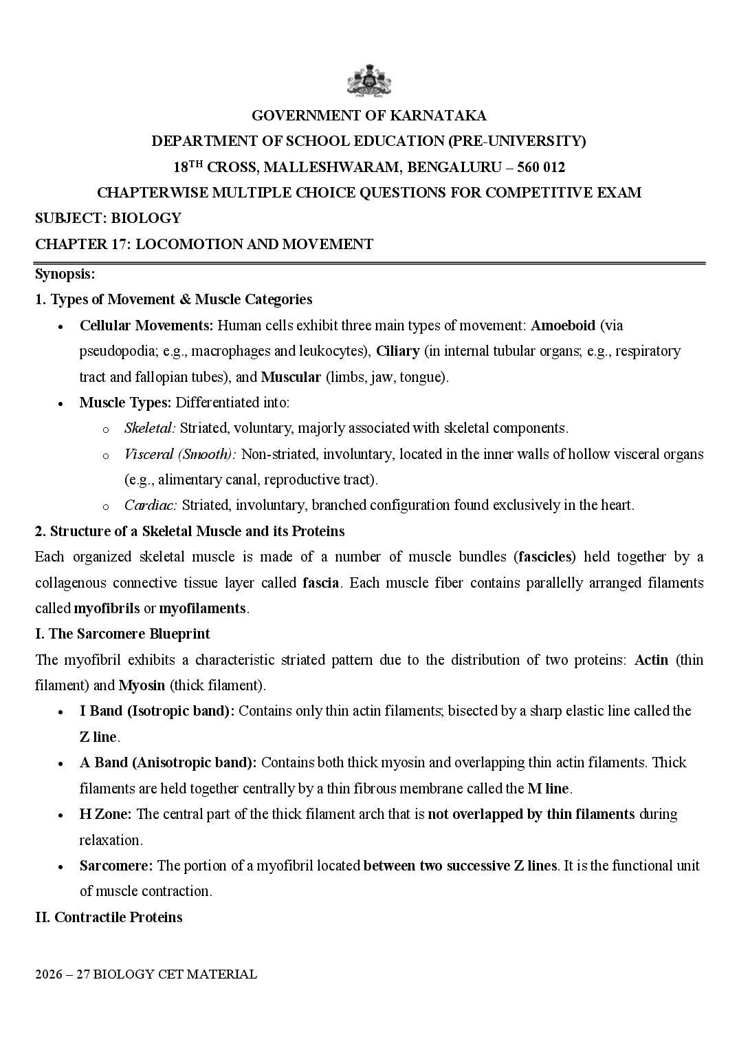
DEPARTMENT OF SCHOOL EDUCATION (PRE-UNIVERSITY)
18TH CROSS, MALLESHWARAM, BENGALURU – 560 012
CHAPTERWISE MULTIPLE CHOICE QUESTIONS FOR COMPETITIVE EXAM
SUBJECT: BIOLOGY
CHAPTER 17: LOCOMOTION AND MOVEMENT
Synopsis:
1. Types of Movement & Muscle Categories
• Cellular Movements: Human cells exhibit three main types of movement: Amoeboid (via
pseudopodia; e.g., macrophages and leukocytes), Ciliary (in internal tubular organs; e.g., respiratory
tract and fallopian tubes), and Muscular (limbs, jaw, tongue).
• Muscle Types: Differentiated into:
o Skeletal: Striated, voluntary, majorly associated with skeletal components.
o Visceral (Smooth): Non-striated, involuntary, located in the inner walls of hollow visceral organs
(e.g., alimentary canal, reproductive tract).
o Cardiac: Striated, involuntary, branched configuration found exclusively in the heart.
2. Structure of a Skeletal Muscle and its Proteins
Each organized skeletal muscle is made of a number of muscle bundles (fascicles) held together by a
collagenous connective tissue layer called fascia. Each muscle fiber contains parallelly arranged filaments
called myofibrils or myofilaments.
I. The Sarcomere Blueprint
The myofibril exhibits a characteristic striated pattern due to the distribution of two proteins: Actin (thin
filament) and Myosin (thick filament).
• I Band (Isotropic band): Contains only thin actin filaments; bisected by a sharp elastic line called the
Z line.
• A Band (Anisotropic band): Contains both thick myosin and overlapping thin actin filaments. Thick
filaments are held together centrally by a thin fibrous membrane called the M line.
• H Zone: The central part of the thick filament arch that is not overlapped by thin filaments during
relaxation.
• Sarcomere: The portion of a myofibril located between two successive Z lines. It is the functional unit
of muscle contraction.
II. Contractile Proteins
Page 2
F-actin is a polymer of monomeric G (globular) actins. Two strands of another protein, Tropomyosin,
run close to the F-actin strands. A complex protein, Troponin, is distributed at regular intervals on the
tropomyosin. In a resting state, a subunit of troponin masks the active binding sites for myosin on the
actin filaments.
• Thick Filament (Myosin): A polymer of monomeric proteins called Meromyosins. Each meromyosin
has two important parts: a Heavy Meromyosin (HMM) (globular head + short arm projecting
outwards) and a Light Meromyosin (LMM) (tail). The globular head contains an Active ATPase
enzyme and has distinct binding sites for ATP and active sites for actin.
3. Mechanism of Muscle Contraction: Sliding Filament Theory
Contraction is initiated by a signal sent by the central nervous system (CNS) via a motor neuron. The motor
neuron plus the muscle fibers it innervates forms a motor unit. The junction between the neuron and the
sarcolemma of the muscle fiber is the Neuromuscular Junction (NMJ) or motor end-plate.
1.Neurotransmitter Release:Action Potential generation.
An action potential reaching the NMJ releases Acetylcholine, which generates a wave of electrical
depolarization across the sarcolemma.
2.Calcium Surge:Sarcoplasmic Reticulum activation.
The action potential spreads through the muscle fiber, driving the sarcoplasmic reticulum to release vast
amounts of Calcium ions ( ) into the sarcoplasm.
3.Unmasking Active Sites: Troponin binding.
Released binds to the -binding subunit of Troponin, inducing a conformational shift that pulls
tropomyosin away, unmasking the active binding sites on actin.
4.Cross-Bridge Formation: ATP Hydrolysis.
Using energy derived from ATP hydrolysis ( ), the energized myosin head binds to the
exposed active sites on actin to form a cross-bridge.
5.The Power Stroke: Sliding inward.
The myosin head rotates, pulling the attached actin filaments inward towards the center of the A band. This
pulls the Z lines closer together, shortening the sarcomere (contraction).
6.Cross-Bridge Breaking: ATP resetting.
A new ATP molecule binds to the myosin head, causing it to detach from actin. The ATP is hydrolyzed once
again to return the myosin head to its high-energy state for the next cycle.
Structural Changes During Contraction Summary:
• The Sarcomere shortens.
• The I bands reduce in width.
Page 3
• The A bands maintain their original length exactly.
• Red vs. White Muscle Fibres:
o Red Fibres (Aerobic): Contain high amounts of the oxygen-storing pigment Myoglobin, a
massive number of mitochondria, and generate ATP via aerobic respiration. They can sustain
contraction for long periods without fatigue.
o White Fibres (Anaerobic): Contain very little myoglobin, fewer mitochondria, but possess a
highly abundant sarcoplasmic reticulum. They depend on anaerobic glycolysis, accumulating
lactic acid quickly, leading to rapid muscle fatigue.
4. The Human Skeletal System (206 Bones)
Divided systematically into the Axial Skeleton (80 bones) and the Appendicular Skeleton (126 bones).
I. Axial Skeleton (80 Bones)
Distributed along the main longitudinal axis of the body:
• Skull (22 bones): Formed of Cranial bones (8) and Facial bones (14). Associated bones include a U-
shaped Hyoid bone (1) at the base of the buccal cavity and Ear Ossicles ( ): Malleus, Incus,
and Stapes.
• Vertebral Column (26 serially arranged vertebrae): Connected dorsally. Formula:
(7 Cervical including Atlas and Axis, 12 Thoracic, 5 Lumbar, 1 fused Sacral, 1 fused Coccygeal/tail
bone).
• Sternum (1): Flat bone on the ventral midline of the thorax.
• Ribs (12 pairs = 24 bones): Bicephalic (possess two articulation facets dorsally).
o True Ribs (1st to 7th pairs): Attached dorsally to vertebrae and ventrally to the sternum via
hyaline cartilage.
o Vertebrochondral / False Ribs (8th, 9th, and 10th pairs): Do not attach directly to the sternum;
join the 7th rib via cartilage.
o Floating Ribs (11th and 12th pairs): Ventrally free, unattached.
II. Appendicular Skeleton (126 Bones)
Comprises the limbs and their supporting girdles:
• Limb Bones ( bones):
o Forelimb (Arm): Humerus (upper arm), Radius and Ulna (forearm), Carpals (8 wrist bones),
Metacarpals (5 palm bones), and Phalanges (14 digits).
o Hindlimb (Leg): Femur (thigh bone - longest/heaviest), Tibia and Fibula, Patella (knee cap),
Tarsals (7 ankle bones), Metatarsals (5 bones), and Phalanges (14 digits).
• Girdles (6 bones): Help articulate the limbs to the axial skeleton.
Page 4
and a large triangular Scapula. The scapula has a sharp spine elevating as the acromion process,
beneath which lies the glenoid cavity for articulation with the humerus head.
o Pelvic Girdle (2 bones): Consists of two coxal bones. Each coxal bone is formed by the fusion
of three bones: Ilium, Ischium, and Pubis. At their point of fusion is a deep cavity called the
acetabulum for articulation with the femur head. The two halves meet ventrally to form the
pubic symphysis containing fibrous cartilage.
5. Joints and Skeletal Disorders
Joints are points of contact between bones, or between bones and cartilages.
• Fibrous Joints: Synarthroses (immovable). Bones are fused end-to-end by dense fibrous connective
tissues (e.g., flat skull bones forming sutures).
• Cartilaginous Joints: Amphiarthroses (limited movement). Bones are joined together via cartilage
bands (e.g., joints between adjacent vertebrae).
• Synovial Joints: Diarthroses (freely movable). Characterized by the presence of a fluid-filled synovial
cavity between the articulating surfaces.
o Ball and Socket Joint: Humerus and pectoral girdle; Femur and acetabulum.
o Hinge Joint: Knee joint, Elbow joint.
o Pivot Joint: Between Atlas and Axis vertebrae.
o Gliding Joint: Between individual Carpal bones.
o Saddle Joint: Between carpal and metacarpal of the thumb.
Disorders of the Muscular and Skeletal System
• Myasthenia Gravis: An autoimmune disorder affecting the neuromuscular junction, leading to fatigue,
weakening, and paralysis of skeletal muscle.
• Muscular Dystrophy: Progressive degeneration of skeletal muscle, mostly due to a genetic disorder.
• Tetany: Rapid spasms (wild contractions) in muscle due to low in body fluid.
• Arthritis: Inflammation of joints.
• Osteoporosis: Age-related disorder characterized by decreased bone mass and increased chances of
fractures. Decreased levels of estrogen is a common cause in aging females.
• Gout: Inflammation of joints due to the accumulation of uric acid crystals.
CLASSROOM DISCUSSION QUESTIONS:
1. In an adult human, how many bones are present as ear ossicles?
(A) 4 (B) 6 (C) 3 (D) None of these
2. The cervical vertebra called axis provides head with sideways rotation. This can be because
(A) It is articulated to skull through occipital condyles
Page 5
(C) It is joined through elastic pads of fibrocartilage with other vertebrae, which provide mobility
(D) It contains odontoid process that fits into the odontoid canal of atlas
3. The given figure is related with myosin monomer (meromyosin). Identify the parts labelled from A to C and
select the correct option.
(A) Head Cross arm GTP binding sites (B) Cross arm Head Ca+2 binding sites
(C) Head Cross arm ATP binding sites (D) Cross arm Head ATP binding sites
4. Humerus with its rounded upper end (head) articulates into
(A) Acromion process (B) deltoid cavity (C) glenoid cavity (D) acetabulum
5. Smallest bone in human system is
(A) Stapes (B) patella (C) malleus (D) incus
6. Collar bone is known as
(A) Scapula (B) clavicle (C) pelvic girdle (D) chevron bone
7. The accumulation of uric acid crystals in the region of joints resulting in painful movements causes
(A) Fluorodosis (B) gout (C) arthritis (D) rheumatoid arthritis
8. Consider the following statements each with one or two blanks.
(i) Repeated activation of the muscles can lead to the accumulation of (1) due to anaerobic breakdown of
glycogen in them, causing fatigue.
(ii) The globular head of meromyosin is an active ATPase enzyme and has binding sites for (2) and active
sites for (3)
(iii) This central part of thick filament, not overlapped by thin filaments is called the (4)
Which one of the following options correctly fills the concerned blanks?
(A) (1) - glucose, (4) - A band (B) (1) - pyruvic acid, (2) - troponin, (3) - myosin
(C) (2) - ATP, (3) - actin, (4) - H zone (D) (1) - lactic acid, (4) - I band
9. Acromian process is characteristically found in the_______ of mammals.
(A) Pectoral girdle (B) sperm (C) pelvic girdle (D) skull
10. The joint in which one of the two bones is fixed in its place and bears a peg like process over which the
other bone rotates is called
(A) Hinge joint (B) saddle joint (C) pivot joint (D) angular joint
11. The slow twitch muscle fibres which are rich in myoglobin and have abundant mitochondria are
Page 6
(C) Red skeletal muscles (D) involuntary muscles
12. The figures given here represent three different conditions of sarcomeres. Identify these conditions and
select the correct option.
A B C
(A) Contracting Relaxed Maximally contracted
(B) Relaxed Contracting Maximally contracted
(C) Maximally contracted Contracting Relaxed
(D) Relaxed Contracted Contracting
13. The type of joint between the human skull bones is called
(A) Cartilaginous joint (B) hinge joint
(C) fibrous joint (D) synovial joint
14. Which of the following components is a part of the pectoral girdle?
(A) Sternum (B) Acetabulum (C) Glenoid cavity (D) Ilium
15. Total number of bones in the hind limb of a man is
(A) 24 (B) 30 (C) 14 (D) 21
16. Match Column - I with Column - II and select the correct option from the codes given below.
Column-I Column-II
(a) Humerus (i) Thigh
(b) Hydrostatic (ii) Upper arm
skeleton
(c) Femur (iii) Flatworms
(iv) Acetabulum
(v) Glenoid
cavity
Page 7
(A) (a) - (ii), (v); (b) - (iii), (vi); (c) - (i), (iv) (B) (a) - (ii), (iv); (b) - (iii), (vi); (c) - (i), (v)
(C) (a) - (i), (v); (b) - (ii), (iv); (c) - (iii), (vi) (D) (a) - (iii), (vi); (b) - (i), (v); (c) - (ii), (iv)
17. Which of the following is the contractile protein of a muscle?
(A) Tropomyosin (B) Tubulin (C) Myosin (D) All of these
18. The functional unit of contractile system in striated muscle is
(A) Sarcomere (B) Z-band (C) cross bridges (D) myofibril
19. The joint between atlas and axis is called
(A) Angular joint (B) hinge joint (C) pivot joint (D) saddle joint
20. Match Column - I with Column - II and select the correct option from the codes given below.
Column-I Column-II
(a) Smooth (i) Myoglobin
muscle
(b) Tropomyosin (ii) Third class
lever
(c) Red muscle (iii) Thin filament
(d) Skull (iv) Sutures
(v) Involuntary
(A) (a) → (iv), (b) → (ii), (c) → (i), (d) → (iii) (B) (a) → (ii), (b) → (iv), (c) → (iii), (d) → (i)
(C) (a) → (iii), (b) → (i), (c) → (iv), (d) → (ii) (D) (a) → (i), (b) → (iv), (c) → (ii), (d) → (iii)
21. What is sarcomere?
(A) Part between two H-lines (B) Part between two A-lines
(C) Part between two I-bands (D) Part between two Z-lines
22. Which of the following pairs is correctly matched?
(A) Hinge joint – Between vertebrae
(B) Gliding joint – Between zygapophyses of the successive vertebrae
(C) Cartilaginous joint – Skull bones
(D) Fibrous joint – Between phalanges
23. Given below is the figure of a sarcomere. Identify the parts labelled as A to D and select the correct option.
Page 8
(A) A band Z line H zone I band
(B) A band H line Z zone I band
(C) I band H line Z zone A band
(D) I band Z line H zone A band
24. During muscle contraction, actin and myosin form
(A) Actomyosin (B) Actoplasm (C) Plastosine (D) Myoplasm
25. The total number of vertebrae in man is
(A) 30 (B) 31 (C) 32 (D) 33
26. The first vertebra, the atlas, by its articulation with axis vertebra facilitates a
(A) Nodding movement (B) Sideway movement
(C) Rotatory movement (D) Backward movement
27. The opening at the base of the skull for the spinal cord is called
(A) Foramen Magnum (B) Foramen of Monro
(B) Obturator foramen (D) Foramen of Magendie
28. What is correct about human body?
(A) There are 5 vertebra in the neck. (B) Brain box is made up of 4 bones.
(C) There are 15 pairs of ribs. (D) There are 12 thoracic vertebra.
29. In mammals, the zygomatic arch is formed by
(A) Maxilla, premaxilla and squamosal. (B) Periotic, jugal and palatine.
(C) Maxilla, squamosal and jugal. (D) Jugal, maxilla and periodic.
30. The last two pairs of ribs are named floating ribs because
(A) Their sterna parts are attached to the sternum directly.
(B) Their sterna parts are attached on the 7 th pair of ribs.
(C) Their sterna parts remain free and do not even reach the sternum.
(D) They float in the body cavity.
31. Vertebral formula for human beings is
(A) C5T12L7S5C3-5 = 33 – 55 (B) C7T12L5S5C3-5 = 33 – 35
(C) C5T10L5S5C3-5 = 33 (D) C7T10L5S5C3–5 = 33
32. How many ribs are present in human beings?
(A) 6 pairs (B) 9 pairs (C) 12 pairs (D) 15 pairs
33. The lower jaw of a mammal is made up
(A) Dentary (B) Maxilla
(C) Premaxilla (D) Post maxilla
34. The cup - shaped structure of pelvic girdle, the acetabulum in man is formed by
Page 9
(C) Ilium, and ischium. (D) Ilium and cotyloid.
35. The articulation of vertebrae in vertebral column occurs when
(A) Post - zygapophyses of a vertebra in front fit beneath the pre - zygapophyses of the another vertebra
behind.
(B) Post - zygapophyses of a vertebra in front fit over the pre - zygapophyses of another vertebra behind.
(C) Pre and post - zygapophyses of a vertebra fit over the post zygapophyses of the verterba in fornt.
(D) Pre and post - zygapophyses of vertebra simply touch one another.
36. Phalangeal formula of hand of man is
(A) 1, 2, 2, 2, 2 (B) 2, 1,1, 1, 1 (C) 2, 3, 3, 3, 3 (D) 2, 3, 3, 2, 2
37. Structure responsible for formation of sigmoid notch is
(A) Olecranon process of humerus. (B) Olecranon process of femur.
(C) Olecranon process of radius ulna. (D) Olecranon process of tibia fibula.
38. Haversian canals are found in
(A) Spinal cord (B) Brain (C) Long bones (D) Sponge
39. Hinge joint is present between
(A) Humerus and pectoral girdle. (B) Femur and acetabulum.
(C) Humerus and radio - ulna. (D)Femur and pelvic girdle.
40. Sutural joints are found between
(A) Parietals of skull (B) Humerus and radio - ulna.
(C) Glenoid cavity and pectoral girdle. (D) Thumb and metatarsal.
41. Which of the following pairs is correctly matched?
(A) Hinge joint Between vertebrae
(B) Gliding joint Between zygapophyses of the successive
vertebrae
(C) Cartilaginous Skull bones
joint
(D) Fibrous joint Between phalanges
42. The contraction of muscle of shortest duration is seen in
(A) Heart (B) Jaws (C) Intestine (D) Eyelids
43. What is sprain?
(A) More pulling of tendon (B) Less pulling of tendon.
(C) More pulling of ligament. (D) Less pulling of ligament.
44. Muscles are red because of the presence of
(A) Myoglobin and mitochondria (B) Haemoglobin and Golgi bodies
Page 10
45. An example of unstriated muscles which are entirely involuntary are located
(A) In the diaphragm (B) In the eyelid
(C) At the pylorus (D) At the base of external ear
46. Electron microscopic studies of the sarcomeres have revealed that during muscle contraction
(A) The width of A – band remains constant. (B) The width of the H – zone becomes smaller.
(C) The width of I – band increases. (D) The diameter of the fibre increases.
47. Which one of the following events is thought to be most closely related to the sliding process between the
two types of filaments which bring about contraction of the fibril?
(A) The liberation of acetyl choline at the motor end plates.
(B) Splitting of an ATP myosin complex.
(C) The influx of sodium ions through the sarcolemma.
(D) Binding of calcium ions.
48. Striated muscles contract by
(A) Sliding of actin filament upon myosin filaments.
(B) Sliding of myosin filaments upon actin filaments.
(C) Pulling together of myosin filaments.
(D) Pulling together of actin filaments.
49. A ……. muscle rotates the forearm to turn the palm downwards.
(A) Pronator (B) Supinator (C) Adductor (D) Adductor
50. In muscular hypertrophy
(A) Cells increase in size by synthesizing the length of a myofibril.
(B) Cells multiply in number.
(C) Cells decrease in size.
(D) Old cells are damaged.
51. The zygomatic process in mammals arises from
(A) Premaxilla (B) Maxilla (C) Mandible (D) Squamosal
52. Bone is distinguished from cartilage by the presence of
(A) Collagen (B) Blood vessels
(C) Lymph vessels (D) Haversian canals
53. Which one of the following is a viral disease that weakens the muscles?
(A) Atrophy (B) Poliomyelitis
(C) Dystrophy (D) Duchene dystrophy
54. Smallest muscle in the human body is
(A) Sartorius (B) Spinal muscle (C) Stapes (D) Stapedius
Page 11
option.
(i)
(ii)
(iii)
(A) (i) – Troponin, (ii) –Tropomyosin, (iii) –F-actin
(B) (i) –Thick filament, (ii) –Troponin, (iii) –Tropomyosin
(C) (i) – Myosin filament, (ii) – Troponin, (iii) – Tropomyosin
(D) (i)–Meromyosin, (ii) – Troponin, (iii) – Tropomyosin
56. Refer to the given figure of a bone. Identify it and choose the correct match for bone and its characteristic
feature from the options given below.
Bone Characteristic Feature
(A) Parietal Protects and supports the throat
(B) Lacrimal Bone of face that articulates with occipital
(C) Hyoid Serves as a point of attachment for some of the muscles of the tongue and
floor of the mouth
(D) Sphenoid Possesses mastoid process
57. Match the following Columns and choose the correct option from the codes given below.
Column I Column II
(a) 1 Axis
(b) 2 Atlas
(c) 3 Thoracic vertebra
Page 12
vertebra
Codes
(a) (b) (c) (d)
(A) 2 4 1 3
(B) 1 2 3 4
(C) 4 3 1 2
(D) 2 1 4 3
58. Refer to the given figure showing enzymatic cleavage of myosin. Read the following statements about it
and choose the option that correctly identifies the true (T) and false (F) statements.
I. Enzyme trypsin splits myosin into two fragments, called Light Meromyosin (LMM) and Heavy
Meromyosin (HMM)
II. HMM lacks adenosine triphosphate activity and does not combine with actin.
III. LMM consists of two globular sub fragment and one rod – shaped fibrous sub fragment.
IV. Each globular sub fragment contains an ATP binding site and actin binding site.
I II III IV
(A) T T F F
(B) T F T F
(C) T F F T
(D) F T T F
59. Given figures are showing the different events of cross – bridge formation during muscle contraction.
Identify them and choose the option that correctly identifies the sequence of their occurrence.
Page 13
60. The given figure is of ribs and rib cage. Choose the incorrect statement about it.
(A) The first 7 pairs of ribs are called true ribs
(B) The 9th, 10th and 11th pairs of the ribs are called false ribs
(C) The last 2 pairs of ribs are called floating ribs
(D) All the statements are incorrect
61. The given figure shows the structure of a sarcomere. Identify the parts labeled as A, B, C and D and choose
the correct option from the codes given below.
A B C D
(A) I– band A –band H–zone M–line
(B) A – band I –band H–zone M–line
(C) I –band A – band M–line Z – line
(D) A – band I –band H–zone M–line
Practice questions
1. Match the following and mark the correct option
Column I Column II
(a) Fast muscle fibres (i) Myoglobin
(b) Slow muscle fibres (ii) Lactic acid
(c) Actin filament (iii) Contractile
unit
(d) Sarcomere (iv) I-band
(A) (a) → (i); (b) → (ii); (c) → (iv); (d) → (iii) (B) (a) → (i); (b) → (ii); (c) → (iii); (d) → (iv)
Page 14
2. Ribs are attached to
(A) Scapula (B) Sternum (C) Clavicle (D) llium
3. What is the type of movable joint present between the atlas and axis?
(A) Pivot (B) Saddle (C) Hinge (D) Gliding
4. ATPase of the muscle is located in
(A) Actinin (B) Troponin (C) Myosin (D) Actin
5. Intervertebral disc is found in the vertebral column of
(A)Birds (B) Reptiles (C) Mammals (D) Amphibians
6. Which one of the following is showing the correct sequential order of vertebrae in the vertebral column of
human beings?
(A) Cervical → lumbar → thoracic → sacral → coccygeal
(B) Cervical → thoracic → sacral → lumbar → coccygeal
(C) Cervical → sacral → thoracic → lumbar → coccygeal
(D) Cervical → thoracic → lumbar → sacral → coccygeal
7. Which one of the following options is incorrect?
(A)Hinge joint− between Humerus and pectoral girdle
(B) Pivot joint− between atlas, axis and occipital condyle
(C) Gliding joint− between the carpals
(D) Saddle joint− between carpel and metacarpals of thumb
8. Knee join and elbow joints are examples
(A) Saddle joint (B) Ball and socket joint
(C) Pivot joint (D) Hinge joint
9. Macrophages and leucocytes exhibit
(A) Ciliary movement (B) Flagellar movement
(C) Amoeboid movement (D) Gliding movement
10. Which one of the following is not a disorder of bone?
(A) Arthritis (B) Osteoporosis (C) Rickets (D) Atherosclerosis
11. Which one of the following statement is incorrect?
(A) Heart muscles are striated and involuntary
(B) The muscles of hands and legs are striated and voluntary
(C) The muscles located in the inner walls of alimentary canal are striated and involuntary
(D) Muscles located in the reproductive tracts are unstriated and involuntary
12. Which one of the following statements is true:
Page 15
(B) Head of humerus bone articulates with glenoid cavity of pectoral girdle.
(C) Head of humerus bone articulates with a cavity called acetabulum of pelvic girdle.
(D) Head of humerus bone articulates with a glenoid cavity of pelvic girdle.
13. Muscles with characteristic striations and involuntary are
(A) Muscles in the wall of alimentary canal (B) Muscles of the heart
(C) Muscles assisting locomotion (D) Muscles of the eyelids
14. Match the followings and mark the correct option
Column I Column II
(a) Sternum (i) Synovial fluid
(b) Glenoid Cavity (ii) Vertebrae
(c) Freely movable (iii) Pectoral
your girdle
(d) Cartilaginous joint (iv) Flat bones
(A) (a) → (ii); (b) → (i); (c) → (iii); (d) → (iv) (B) (a) → (iv); (b) → (iii); (c) → (i); (d) → (ii)
(C) (a) → (ii); (b) → (i); (c) → (iv); (d) → (iii) (D) (a) → (iv); (b) → (i); (c) → (ii); (d) → (iv)
Assertion and Reason type questions
In the following questions, a statement of Assertion (A) is followed by a statement of Reason (R). Mark the
correct choice as :
(A) If both Assertion and Reason are true and Reason is the correct explanation of Assertion
(B) If both Assertion and Reason are true but Reason is not the correct explanation of Assertion
(C) If Assertion is true but Reason is false
(D) If both Assertion and Reason are false.
15. Assertion: Locomotion enables the animal to find its partner.
Reason: Locomotion enables the animal to shift its entire body from place to place.
16. Assertion: Movement of body parts serves to change the body posture.
Reason: Body parts move in relation to body axis.
17. Assertion: Skeletal muscles become stiff after death.
Reason: The production of ATP ceases and the proteins of muscle fibres become irreversibly bound
together.
18. Assertion: There are similarities between the locomotion of unicellular organism and multicellular animal.
Reason: Ciliary, flagellar and amoeboid movement occur in unicellular organisms.
19. Assertion: Locomotion in Hydra is carried out by two types of contractile cells.
Reason: Muscle fibres are lacking in Hydra.
Page 16
Reason: Starfish utilise a flow of water over the surface, for movement.
21. Assertion: Skeleton serves as a storage depot.
Reason: Skeleton stores carbohydrate and protein.
22. Assertion: Ball and - socket - joints are the most mobile joints.
Reason: Synovial fluid is present here.
23. Assertion: Arthritis or inflammation of a joint makes the joint painful.
Reason: Some toxic substances are deposited at the joint.
24. Assertion: Movement and locomotion are indispensable for all the vital activities among animals.
Reason: Locomotion distinguishes most animals from plants.
25. Assertion: The contraction and relaxation of muscle fibre is controlled by nerve impulses.
Reason: The threshold stimulus is the minimum stimulus required for the beginning of contraction.
26. Assertion: Muscle twitch is related with tetanus.
Reason: Tetanus is caused by many nerve impulses at a time.
27. Assertion: Extra oxygen consumption in human body is known as oxygen debt.
Reason: The extra oxygen is required by the body to oxidise the accumulated lactic acid produced
during strenous exercise.
28. Assertion: Athletic training enhances the capacity for aerobic contraction of muscles.
Reason: After strenous exercise, athletes are liable to much more oxygen debt than non-athlete.
29. Assertion: The five lower pairs of ribs in man are known as false ribs.
Reason: They are attached behind to the thoracic vertebrae but in front not attached directly to the
breastbone.
30. Assertion: Hydra can exhibit a variety of locomotory movements.
Reason: Contraction and relaxation of muscle fibres cause all sorts of movements and loco-motion in
Hydra.
31. Assertion: Triceps is said to be an extensor muscle for elbow joint.
Reason: Triceps relaxes during extension of forearm at the elbow joint.
32. Assertion: Biceps and triceps are called antagonistic muscles.
Reason: This is due to the fact that they contract and relax together.
33. Assertion: In each sarcomere, the thick filaments are present at the two ends, whereas the thin filaments are
located centrally.
Reason: The thick and thin filaments are alternately arranged and they don't overlap.
34. Assertion: In arthritis, the movements at the joints may be immobilised.
Reason: Any mechanical injury and defects in ligaments can cause displacement of bones from their
normal position.
Page 17
Reason: This is due to increased contraction of individual muscle fibres with increase in stimulus strength.
36. Assertion: Earthworm and leeches have longitudinal and circular muscle fibres in their body wall.
Reason: These animals do not possess any skeletal system.
37. Assertion: Muscle as a whole doesn't obey All or None law.
Reason: Each muscle fibre contracts maximally whenever it contracts.
38. Assertion: Biceps is said to be a flexor muscle for elbow joint.
Reason: Biceps contract during flexion at the elbow joint.
39. Assertion: The synaptic channels on the end plate of skeletal muscle are activated by atropine.
Reason: Atropine is a neurotransmitter.
40. Assertion: During normal movement slow twitch muscle fibres are recruited first.
Reason: Slow twitch muscle fibres have low myosin-adenosine triphosphatase (ATPase) activity.
41. Assertion: Little rotation and bending of the I. vertebral column is possible.
Reason: This is due to the presence of fibrous joints between vertebrae.
42. Assertion: During contraction, neither the thin filaments nor the thick filaments change in length when the
muscle contracts.
Reason: The filaments slide past each other longitudinally, so that the degree of overlap of the thin and
thick filaments increases.
43. Assertion: White muscles depend mainly on anaerobic glycolysis for energy production.
Reason: White muscles carry out anaerobic contractions, accumulate malic acid in considerable amounts
during strenuous work and soon get fatigued.
44. Assertion: The red muscle fibres can go on contracting for prolonged durations without fatigue.
Reason: The red muscles contain very high amount of myoglobin.
45. Refer to the given figure of lumbar vertebra of man. Identify the parts labelled as A-D and choose the
correct option from the codes given below.
A B C D
(A) Transverse Superior Vertebral Spinous
Process Articular process Foramen Process
(B) Spinous Superior articular Vertebral Transverse
Process process Foramen Process
Page 18
Process foramen Articular Process
Process
(D) Transverse Vertebral Superior Spinous
Process foramen Articular Process
Process
46. Consider the following figure of motor unit. Read the following statements about it and choose the correct
option from the codes given below.
I. A neuron that transmits a stimulus to muscle tissue is called motor neuron.
II. A motor unit consists of two adjacent motor neurons and the muscle fibres it innervates.
III. The portion of the muscle plasma membrane that lies beneath the nerve endings is called the motor end
plate.
IV. The axon terminals and the motor end plate together constitute the neuromuscular junction.
(A) I and II (B) II and III
(C) I, II and III (D) I, III and IV
47. Refer to the given figure of a meromyosin. Read the following statements about it and choose the correct
option.
I. Myosin contributes 55% of muscle protein by weight.
II. The myosin molecule is composed of six polypeptide chains, two identical light chains and four heavy
chains.
III. The heavy chains wrap spirally around each other to form a double helix.
IV. One end of each of these chains is folded into a globular protein mass called myosin head.’
Choose the correct option.
(A) I, III and IV (B) II and III
(C) I, II and III (D) I, II, III and IV
Page 19
functions of its different parts, read them and choose the correct option.
I. B is a membrane composed of secretory epithelial cells which secretes a thick sticky fluid.
II. C acts as a lubricant to joint, provides nutrient materials for the structures within the joint cavity and
helps to maintain the stability of the joint.
III. D is composed of hyaline cartilage.
IV. A joins the bones.
(A) I and II (B) III and IV
(C) I and IV (D) All of the above
49. Match the following columns and choose the correct option from the codes given below.
Column I Column II
(a) 1 partially contracted muscle
(b) 2 relaxed muscle
(c) 3 maximally contracted
muscle
Codes
(a) (b) (c)
(A) 1 2 3
(B) 3 2 1
Page 20
(D) 2 1 3
50. The given figure is showing pelvic girdle and bones of legs. Observe it carefully and identify the parts
labeled as A, B, C, D and E.
A B C D E
(A) Ischium Ilium Fibula Tibia Patella
(B) Ilium Ischium Fibula Tibia Condyle
(C) Ilium Ischium Tibia Fibula Patella
Pubic
(D) Sacrum Tibia Fibula Condyle
symphysis
51. Identify A, B, C and D in the given diagram of human skull. Choose the correct option.
A B C D
(A) Hyoid bone Maxilla Frontal bone Parietal bone
(B) Hyoid bone Maxilla Parietal bone Frontal bone
(C) Maxilla Hyoid bone Parietal bone Frontal bone
(D) Parietal bone Frontal bone Maxilla Hyoid bone
52. Given figure is of three different proteins of thin myofilaments. Choose the incorrect statement
about them.
Page 21
(B) Tropomyosin is a double stranded – helical rod
(C) Troponin is a complex of three polypeptides; troponin T (TP T), troponin I (TP I), and troponin
C (TP C)
(D) Troponin C(TP C) is a calcium – binding polypeptide
53. Match the following Columns and choose the correct option from the codes given below.
Column I Column II Column III
1 A. Hinge joint (i) Hip joint
2 B. Ball and socket (ii) Between carpal and metacarpal of thumb of
joint hand
3 C. Saddle joint (iii) Between atlas and axis
4 D. Pivot joint (iv) Knee
Codes
1 2 3 4
(A) B, (i) D, (iii) A, (iv) C, (ii)
(B) A, (i) B, (ii) C, (iii) D, (iv)
(C) C, (ii) D, (iv) A, (iii) B, (i)
(D) A, (iii) C, (ii) B, (iv) D, (i)
54. Carefully observe the given diagram showing the antagonistic arrangement of biceps and triceps muscle.
Page 22
II. Triceps are antagonist to biceps, extends the arms and is called an extensor.
III. When a flexor contracts, the opposing extensor must relax to permit the bone to move.
IV. Similar pairs of opposing flexors and extensors are found at wrist, knee and ankle.
Choose the correct option.
(A) I and IV (B) II and III (C) II and IV (D) All of the above
55. The given figure is of a condyloid joint. Choose the incorrect statement about it.
(A) In this type of joint an oval – shaped condyle of one bone fits into an elliptical cavity of another bone
(B) Condyloid joint allows movement in only one plane, i. e. back and forth
(C) The joints between the metacarpals and phalanges of the fingers are examples of condyloid joints
(D) It is a biaxial joint
56. Identify A, B and C in the given diagram and choose the correct option.
(A) A – Cervical vertebrae, B – Coccyx, C – Sacrum, D – Thoracie vertebrae
(B) A – Cervical vertebrae, B – Coocyx, C – Atlas, D – Thoracie vertebrae
(C) A – Cervical vertebrae, B – Coccyx, C – Axis, D– Thoracie vertebrae
(D) A – Cervical vertebrae, B – Sacrum, C – Coccyx, D – Thoracie vertebrae
57. The given figure is showing different types of synovial joints. Identify the joints A – D in the figure.
Page 23
(B) A – Saddle joint, B – Pivot joint, C – Condyloid joint, D – Gliding joint
(C) A – Pivot joint, B – Gliding joint, C – Saddle joint, D – Condyloid joint
(D) A – Pivot joint, B – Saddle joint, C – Gliding joint, D – Condyloid joint
58. The given figure is showing the hands of a patient suffering from gouty arthritis. Choose the incorrect
statement about it.
(A) This disease is due to defect in purine metabolism that causes an excess of uric acid and its salts
(B) Crystals of uric acid salts accumulate in joints
(C) The excess of urates can form stones in gall bladder
(D) Treatment with certain drugs can increase the excretion of urates
59. The given figure is showing the anterior view of human sacrum and coccyx. Choose the incorrect statement
about it.
(A) The five sacral vertebrae fuse to form the sacrum
(B) The female sacrum is shorter, wider than the male sacrum
(C) The four coccygeal vertebrae fuse to form a curved triangular bone, called the coccyx
(D) In females, the coccyx points anteriorly, but in males it points inferiorly
Classroom discussion Keys
Page 24
2. (D) 10. (C) 18. (A) 26. (C) 34. (A) 42. (D) 50. (B) 58. (C)
3. (C) 11. (C) 19. (C) 27. (A) 35. (B) 43. (C) 51. (D) 59. (C)
4. (C) 12. (B) 20. (A) 28. (D) 36. (C) 44. (A) 52. (D) 60. (B)
5. (C) 13. (C) 21. (D) 29. (C) 37. (A) 45. (C) 53. (B) 61. (A)
6. (B) 14. (C) 22. (B) 30. (C) 38. (C) 46. (A) 54. (D)
7. (B) 15. (B) 23. (A) 31. (B) 39. (C) 47. (D) 55. (A)
8. (C) 16. (A) 24. (A) 32. (C) 40. (A) 48. (A) 56. (C)
Practice question Keys
1. (C) 11. (C) 21. (C) 31. (C) 41. (C) 51. (D)
2. (B) 12. (B) 22. (B) 32. (C) 42. (A) 52. (A)
3. (A) 13. (B) 23. (B) 33. (D) 43. (C) 53. (A)
4. (B) 14. (B) 24. (B) 34. (B) 44. (A) 54. (D)
5. (C) 15. (B) 25. (B) 35. (C) 45. (B) 55. (B)
6. (D) 16. (B) 26. (B) 36. (B) 46. (D) 56. (D)
7. (A) 17. (A) 27. (A) 37. (B) 47. (A) 57. (C)
8. (D) 18. (B) 28. (C) 38. (A) 48. (A) 58. (C)
9. (C) 19. (A) 29. (A) 39. (D) 49. (D) 59. (D)
10. (D) 20. (B) 30. (C) 40. (A) 50. (C)
Document Details
| Board / Org | Karnataka Board |
| Exam | Class 11 |
| Type | Question Bank |
| Pages | 24 |
| Updated | 24 Sep 2026 |