HBSE Class 11 Sample Paper 2026 Answers Biotechnology
Finished viewing? Save it for later —
Download HBSE Class 11 Sample Paper 2026 Answers Biotechnology (PDF · 22 pages)About HBSE Class 11 Sample Paper 2026 Answers Biotechnology
HBSE Class 11 Sample Paper 2026 Answers Biotechnology is available here for free download. Published by Haryana Board for Class 11, this answer key can be viewed online or downloaded as a PDF (22 pages). Candidates preparing for Class 11 can use HBSE Class 11 Sample Paper 2026 Answers Biotechnology to understand the exam pattern, the type of questions asked, and the overall difficulty level.
Frequently Asked Questions
How can I download HBSE Class 11 Sample Paper 2026 Answers Biotechnology?
Open this page and click the Download button to save HBSE Class 11 Sample Paper 2026 Answers Biotechnology as a PDF. It is completely free on AglaSem Docs.
Is HBSE Class 11 Sample Paper 2026 Answers Biotechnology free to download?
Yes. HBSE Class 11 Sample Paper 2026 Answers Biotechnology can be viewed online and downloaded as a PDF free of cost on AglaSem Docs.
How many pages does HBSE Class 11 Sample Paper 2026 Answers Biotechnology have?
HBSE Class 11 Sample Paper 2026 Answers Biotechnology contains 22 pages, which you can read online or download together as a single PDF.
Where can I find more Class 11 study material?
You can find more Class 11 question papers, sample papers, syllabus, and answer keys on AglaSem Docs.
HBSE Class 11 Sample Paper 2026 Answers Biotechnology – Text
Read the full text of this answer key below — useful to quickly search, copy and reference the content online without downloading the PDF.
📄 View text version (20 pages)
Page 1
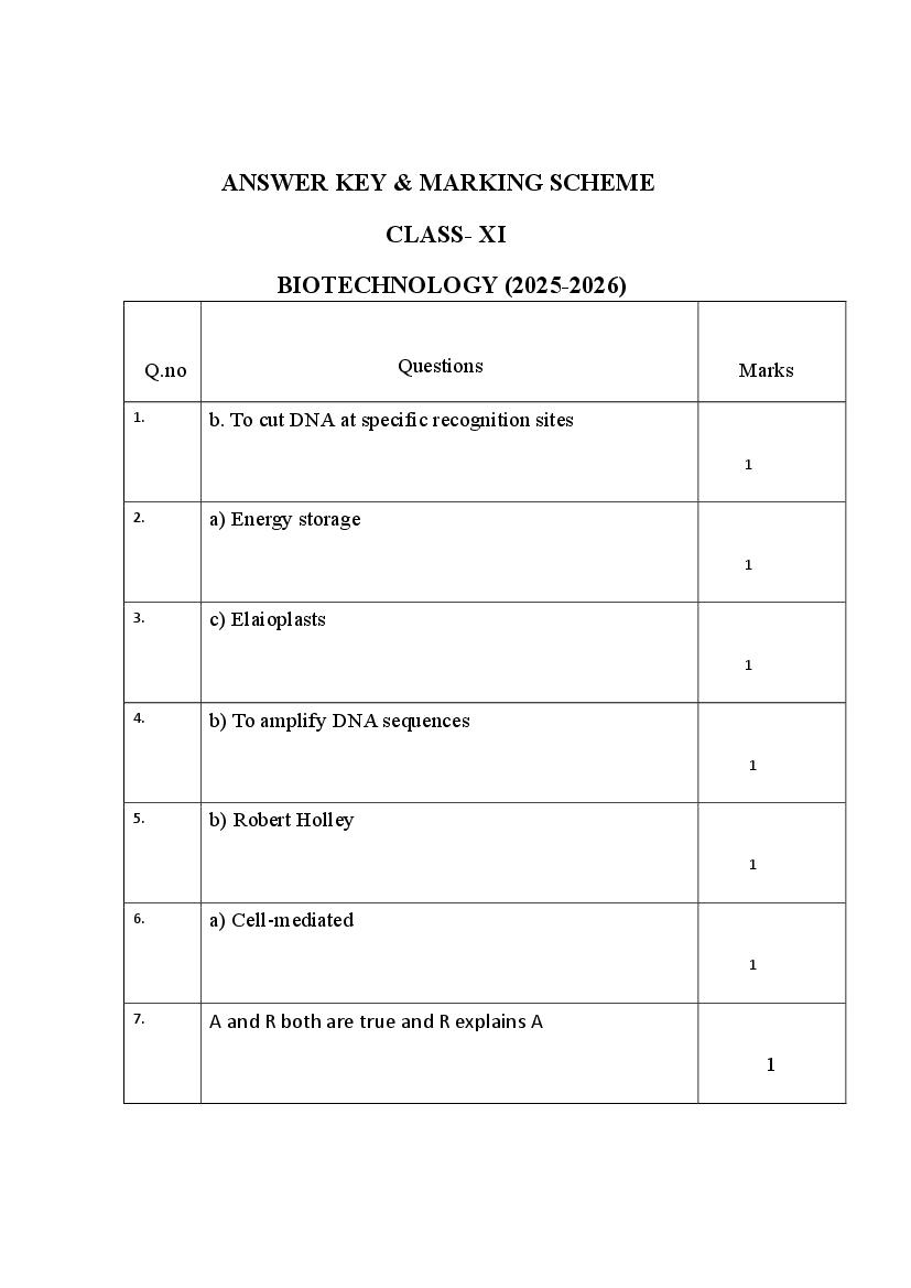
CLASS- XI
BIOTECHNOLOGY (2025-2026)
Q.no Questions Marks
1. b. To cut DNA at specific recognition sites
1
2. a) Energy storage
1
3. c) Elaioplasts
1
4. b) To amplify DNA sequences
1
5. b) Robert Holley
1
6. a) Cell-mediated
1
7. A and R both are true and R explains A
1
Page 2
1
9. A is true, R is false
1
10. Variable Number Tandem Repeats
1
11. Bioreactors
1
12. Ribosome
1
13. Taq polymerase
1
14. Golgi apparatus
1
15.
DNA and proteins
1
Page 3
provide a controlled environment for the growth of
microorganisms, cells, or tissues to produce biological products
like enzymes, vaccines, or recombinant proteins.
They maintain optimal conditions such as temperature, pH,
oxygen supply, and nutrients to support biological processes
efficiently 2
Example: A Stirred Tank Bioreactor is a common example. It is
used for large-scale production of insulin, antibiotics, or
bioenzymes.
17. The structural unit of DNA is called a nucleotide.
1. A nitrogenous base (Adenine, Thymine,
Cytosine, or Guanine)
2. A deoxyribose sugar
3. A phosphate group
Or
Applications of Biotechnology in Medicine:
1. Production of Insulin – Recombinant DNA
technology is used to produce human insulin for
diabetic patients.
2. Gene Therapy – Used to correct genetic disorders
by inserting healthy genes.
2
3. Production of Vaccines – Biotechnology helps in
developing modern vaccines, like Hepatitis B and
COVID-19.
4. Monoclonal Antibodies – Used in diagnosis and
treatment of cancers and autoimmune diseases.
5. Personalized Medicine – Tailored treatments
based on individual genetic makeup.
Page 4
“Every individual possesses two alleles for each trait,
and these alleles separate (segregate) during the
formation of gametes, so that each gamete carries only
one allele for each trait.”
Relation to Mendelian Inheritance:
• This law is the first law of Mendel.
• It explains how traits are inherited from one
generation to the next.
• During meiosis, the two alleles of a gene present
in a parent segregate into different gametes,
ensuring equal chances of passing either allele to 2
offspring.
• Example: In a cross between Tt × Tt, the tall (T)
and short (t) alleles segregate, leading to a 3:1
ratio in F2 generation (Tall : Short)
Page 5
molecules found in bacteria that can replicate
independently of the bacterial chromosome.
When used as vectors in genetic engineering, plasmids
act as carriers to transfer foreign genes into a host cell
(usually bacteria) for cloning or expression of those
genes.
Why Plasmids are Good Vectors:
• They have a replication origin (ori) – so they can
copy themselves. 2
• Can be easily modified in labs.
• Often contain selectable markers like antibiotic
resistance genes.
Page 6
20. and SER (Smooth Endoplasmic Reticulum):
Feature
SER (Smooth
Endoplasmic
RER (Rough Endoplasmic Reticulum)
Reticulum) Absent on surface,
Ribosomes hence smooth
appearance
Present on the surface,
giving it a rough
appearance Synthesis of lipids and
steroids, detoxification
Main of drugs
Synthesis of proteins
Function
Page 7
Appearance Rough and grainy under Smooth and tubular
microscope
Golgi apparatus and
Associated Lipid metabolism and
protein transport
With detoxification processes
Spread throughout the
Common near the cytoplasm
Location
nucleus
Cholesterol, hormones,
membrane
Enzymes, hormones,
Examples of lipids
membrane proteins
Products
Page 8
Or
Structure of Plasma Membrane – Singer & Nicolson Model
(Fluid Mosaic Model)
The Fluid Mosaic Model was proposed by Singer and
Nicolson in 1972. According to this model:
• The plasma membrane is a flexible (fluid) bilayer
phospholipids.
• Proteins are embedded within this bilayer like a
mosaic.
• The membrane is semipermeable, allowing selective
movement of substances.
Page 10
Haemophilia is a genetic disorder in which the blood lacks
certain clotting factors, resulting in delayed or prolonged
bleeding.
• It is a sex-linked recessive disorder, mostly affecting
males.
• Caused due to mutation in genes coding for Factor
VIII or IX.
• Even minor injuries can lead to severe internal
bleeding.
• Inheritance: Carried by females (carriers) and passed 2
on to male children.
Thalassemia
Thalassemia is an inherited blood disorder in which the body
makes an abnormal form or inadequate amount of
haemoglobin.
• This leads to destruction of red blood cells and causes
anaemia.
• Two main types:
o Alpha-thalassemia
o Beta-thalassemia (more common in India)
• Symptoms: Weakness, pale skin, fatigue, delayed
growth.
• Treatment: Regular blood transfusions, iron chelation
therapy, and in severe cases, bone marrow transplant.
Or
Gene Mapping – Explanation
Gene Mapping is the process of determining the specific
location (position) of genes on a chromosome. It helps in
identifying the order and distance between genes based on
how often they are inherited together.
Key Points:
• Shows the linear arrangement of genes.
• Helps in locating disease-causing genes.
• Measured in map units or centimorgans (cM).
Page 12
centimorgan = 1% chance of recombination
between two genes during meiosis.
The concept of gene mapping was first given by Thomas
Hunt Morgan and his student Alfred Sturtevant in the early
1900s while working on Drosophila (fruit fly).
• Alfred Sturtevant is credited with creating the first
genetic map in 1913.
22. The Central Dogma of Molecular Biology describes the flow
of genetic information within a biological system.
The Central Dogma states that:
DNA → RNA → Protein
This means genetic information flows from:
3
1. DNA (Deoxyribonucleic acid) →
2. RNA (Ribonucleic acid) →
3. Protein (functional molecules in the cell)
Steps Involved:
1. Transcription o DNA is used to create a
complementary mRNA strand.
o Occurs in the nucleus.
o Example: A → U, T → A, G → C, C → G
2. Translation o mRNA is read by ribosomes to
assemble amino acids into a protein.
o Occurs in the cytoplasm (at ribosomes).
o Each codon (3 RNA bases) codes for one amino
acid.
Importance:
• Explains how genes control cell function through the
production of proteins.
• Fundamental to understanding genetic expression and
biotechnology.
23. Feature
Plant Cell Animal Cell
Structure
Page 13
Absent
Cell Wall cellulose)
Usually rectangular or Usually round or 3
Shape box-like irregular
Present (for
Absent
Chloroplasts photosynthesis)
Many small vacuoles (or
Vacuole One large central vacuole sometimes none)
Absent (except in lower Present (important in
Centrioles
plants) cell division)
Lysosomes Rare or absent Common
Present (chloroplast,
Not present
Plastids chromoplast, etc.)
Energy Storage Starch Glycogen
Cilia and Often present in some
Rare
Flagella cells (e.g., sperm)
Function of Stores water, maintains Helps in digestion and
Vacuole turgor pressure waste removal
Or
What is Point Mutation?
A point mutation is a type of gene mutation where a single nucleotide
base (A, T, C, or G) in the DNA sequence is changed, inserted, or
deleted.
It affects only one point (base pair) in the DNA sequence — hence the
name point mutation.
Types of Point Mutation:
1. Substitution – One base is replaced by another. Example:
o Normal DNA: A A G (codes for Lysine) o
Mutated DNA: A G G (codes for Arginine)
2. Insertion – An extra base is added.
Example: A A G → A A **T** G (may shift the reading frame)
3. Deletion – A base is removed.
Example: A A G → A _ G (frameshift mutation)
Page 14
• Sickle Cell Anemia:
A single base substitution (A → T) changes the codon from
GAG (Glutamic acid) to GTG (Valine) in the hemoglobin gene.
• Cystic Fibrosis:
Caused by deletion of a single base leading to production of a
non-functional protein.
24. Protein synthesis is the biological process by which cells build
proteins using the instructions from DNA. It involves two main
stages: Transcription and Translation.
3
Step 1: Transcription (DNA → mRNA)
Location: Nucleus
• DNA is used as a template to create messenger RNA (mRNA).
• The enzyme RNA polymerase reads the DNA strand and creates a
complementary mRNA strand.
• Example:
DNA: TAC GGA → mRNA: AUG CCU
This step copies the gene's instructions into a portable message (mRNA).
Step 2: mRNA Leaves Nucleus
• The mRNA exits the nucleus and enters the cytoplasm to find a
ribosome.
Step 3: Translation (mRNA → Protein)
Location: Ribosome (in cytoplasm)
• The ribosome reads the mRNA 3 bases at a time (codon).
• Each codon codes for a specific amino acid.
• tRNA (transfer RNA) brings the correct amino acid to the ribosome.
• The amino acids are linked together to form a protein chain
(polypeptide).
Step 4: Protein Formation
• Once a stop codon is reached, the protein is released and folded into
its functional shape.
Page 15
1. DNA gives code.
2. mRNA carries the code.
3. Ribosome reads the code.
4. tRNA brings amino acids.
5. Amino acids form protein.
25. Feature RER (Rough ER) SER (Smooth ER)
Present on surface
Ribosomes (gives rough appearance)
Absent on surface (smooth
appearance)
Lipid synthesis,
Protein synthesis and detoxification, and hormone
Main Function transport production
Appearance Flattened sacs Tubular structure
Location Near the nucleus Throughout the cytoplasm
Works in metabolic
Associated
Works with Golgi body processes like fat
Organelles for protein modification metabolism
Example of Enzymes, membrane Steroids, lipids, detox
Products proteins enzymes
Endoplasmic
Feature Golgi Complex Reticulum (ER)
Stack of flattened, Network of tubules and
Structure membrane-bound sacs sacs spread throughout
(cisternae) cytoplasm
Two types: RER (with
Types No types ribosomes) and SER
(without ribosomes)
Modifies, sorts, and Synthesizes proteins
Function
packages proteins & lipids (RER) and lipids (SER)
Continuous with the
Location Near the nucleus and ER
nuclear envelope
Involved in
Involvement in Involved in synthesis
shipping/packaging of
Transport and initial transport
cellular products
Page 16
Vesicles secretion Golgi apparatus
Discovery Discovered by Camillo Discovered by Keith
Golgi Porter
26. Complex Tissue in Plants and Their Functions
Name of Complex Tissues:
1. Xylem
2. Phloem
These are called complex tissues because they are made up of more
than one type of cell, working together to perform a specific function.
3
1. Xylem – Water and Mineral Transport Function:
• Conducts water and dissolved minerals from roots to other parts
of the plant.
• Also provides mechanical support.
Cell Types in Xylem:
• Tracheids
• Vessels
• Xylem parenchyma
• Xylem fibres
2. Phloem – Food Transport Function:
• Transports prepared food (mainly sucrose) from leaves to other
parts of the plant (source to sink).
• Supports growth and storage.
Cell Types in Phloem:
• Sieve tubes
• Companion cells
• Phloem parenchyma
• Phloem fibres
27. Who Gave the Concept of Gene Mapping?
The concept of Gene Mapping was first introduced by
Alfred Sturtevant in 1913, a student of Thomas Hunt
Morgan. He worked on fruit flies (Drosophila) and
constructed the first genetic linkage map.
Page 17
Gene mapping is the process of determining the location
and order of genes on a chromosome. It helps in identifying
where a gene is located and how closely two genes are
linked based on recombination frequency.
Unit of Gene Mapping: 3
• The unit used is centiMorgan (cM).
• 1 centiMorgan = 1% recombination frequency
between two genes.
Explanation (Simple Terms):
• Genes that are closer together on a chromosome are
less likely to be separated during recombination
(crossing over).
• The further apart the genes are, the higher the
chance of recombination.
• Gene maps help researchers in:
o Identifying disease-causing genes o
Studying genetic inheritance o Advancing
biotechnology and genomics
28. What is Double Fertilization?
Double fertilization is a unique process found only in
flowering plants (angiosperms) where two male gametes fuse
with two different cells in the embryo sac of the ovule. This
process was discovered by Nawaschin in 1898.
Steps of Double Fertilization: 5
1. Pollination – Pollen lands on the stigma of a flower.
2. Pollen Tube Formation – Grows through the style and
enters the ovule via the micropyle.
3. Two Male Gametes Released into the embryo sac.
4. First Fertilization (Syngamy):
o One male gamete fuses with the egg cell.
Page 18
embryo.
5. Second Fertilization (Triple Fusion):
o Other male gamete fuses with two polar nuclei.
o Forms a triploid (3n) cell → develops into
endosperm (nutritive tissue).
Result of Double Fertilization:
• Zygote (2n) → forms the embryo
• Endosperm (3n) → provides nutrition to the growing
embryo
Why It Is Called "Double"?
Because two fertilizations happen:
1. Egg + Sperm → Zygote
2. Polar nuclei + Sperm → Endosperm
Or
Different Phases of the Cell Cycle – Explained
The cell cycle is the series of events that a cell goes through
to grow and divide. It consists of two main stages:
1. Interphase – Cell prepares for division
2. M Phase (Mitotic phase) – Cell actually divides
1. Interphase (Preparation Phase)
Interphase takes up about 90% of the cell cycle. It has three
sub-phases:
a) G₁ Phase (Gap 1)
• The cell grows in size.
• Proteins and organelles are synthesized.
• The cell performs normal functions.
b) S Phase (Synthesis Phase)
• DNA replication occurs → chromosomes duplicate.
• Each chromosome becomes two sister chromatids.
c) G₂ Phase (Gap 2)
• The cell prepares for mitosis.
• Enzymes and other proteins required for cell division
are produced.
Page 19
This phase includes the division of the nucleus and
cytoplasm. It has two parts:
a) Mitosis (Karyokinesis) – Division of the nucleus
Consists of 4 stages:
1. Prophase – Chromosomes condense, spindle forms.
2. Metaphase – Chromosomes align at the center
(equator).
3. Anaphase – Sister chromatids separate and move to
opposite poles.
4. Telophase – Nuclear membranes reappear,
chromosomes decondense.
b) Cytokinesis – Division of the cytoplasm
• Two daughter cells are formed.
29.
What is Pedigree Analysis?
Pedigree analysis is a diagrammatic method used to study the
inheritance of traits (especially genetic disorders) across
multiple generations of a family.
• It uses symbols to represent family members and
shows how traits or disorders are passed on.
• Commonly used in genetics, medicine, and counseling.
How It Helps in Identifying Genetic Disorders: 5
1. Tracks inheritance patterns of a disease (dominant,
recessive, sex-linked).
2. Helps in identifying carriers of genetic disorders.
3. Predicts the probability of a disorder appearing in
future generations.
4. Distinguishes between:
o Mendelian disorders (like haemophilia,
thalassemia) o Chromosomal disorders (like
Down syndrome)
Symbols Used in Pedigree Charts:
Symbol Meaning
☐ Unaffected male
○ Unaffected female
Page 20
● Affected female
─┬─ Parents connected to children
OR
Mendelian Chromosomal Disorders
Feature
Disorders
Caused by change in
Cause Caused by mutation number or structure of
in a single gene chromosomes
Follows Mendel’s
Inheritance Does not follow
laws (dominant or
Pattern Mendelian inheritance
recessive)
Number of
Multiple genes on the
Genes Usually one gene
affected chromosome(s)
Affected
Detected by Detected by karyotyping
pedigree analysis (chromosome
Diagnosis
and molecular examination)
techniques
- Sickle Cell
- Down Syndrome -
Examples Anemia -
Turner Syndrome -
Thalassemia -
Klinefelter Syndrome
Hemophilia
Page 21
Genotype:
• Definition: The genetic makeup of an organism that
determines a particular trait.
• It refers to the combination of alleles inherited from
the parents.
• Example: o For tallness in pea plants: TT or Tt is the
genotype.
o For dwarf: tt
Genotype is not always visible but controls what the
organism can express.
5
Phenotype:
• Definition: The observable physical traits or
characteristics of an organism.
• It is the expression of the genotype in interaction with
the environment.
• Example: o Tall or short pea plant.
o Brown eyes, curly hair, etc. Phenotype
is what you can see or measure.
Or What
is Haemophilia?
Haemophilia is a genetic disorder in which a person’s blood
lacks clotting factors, making it difficult for blood to clot
after an injury.
• It leads to prolonged bleeding, even from small cuts or
internal injuries.
• People with haemophilia may suffer from spontaneous
bleeding in joints, muscles, and internal organs.
Types of Haemophilia:
1. Haemophilia A – Deficiency of Clotting Factor VIII 2.
Haemophilia B – Deficiency of Clotting Factor IX
Page 22
• It is a sex-linked recessive disorder.
• The gene responsible is present on the X chromosome.
• Males (XY) are more commonly affected.
• Females (XX) are usually carriers and rarely show
symptoms.
Why is it Called “Royal Disease”?
• Haemophilia was famously present in the royal
families of Europe, especially in Queen Victoria’s
descendants.
• Queen Victoria was believed to be a carrier of the
disease and passed it on to several royal families in
Russia, Germany, and Spain.
• Since it spread through royal bloodlines, it became
known as the “Royal Disease.”
Symptoms of Haemophilia:
• Easy bruising
• Excessive bleeding after injuries or surgeries
• Pain and swelling in joints due to internal bleeding
Treatment:
• Replacement therapy: Injecting the missing clotting
factor
• Avoiding injury
• Gene therapy (under research)
Document Details
| Board / Org | Haryana Board |
| Exam | Class 11 |
| Type | Sample Paper |
| Pages | 22 |
| Updated | 24 Sep 2026 |